Microsoft word - 1-capa.doc
Pontifícia Universidade Católica do Rio Grande do Sul
Faculdade de Biociências
Mestrado em Biologia Celular e Molecular - PPGBCM
TRATAMENTO DE ESTRIAS ALBAS COM GALVANOPUNTURA:
BENÉFICO PARA A ESTÉTICA, ESTRESSE OXIDATIVO E PERFIL LIPÍDICO
Shanna Bitencourt
Prof. Dr. Jarbas Rodrigues de Oliveira
Porto Alegre, RS
Pontifícia Universidade Católica do Rio Grande do Sul
Faculdade de Biociências
Mestrado em Biologia Celular e Molecular - PPGBCM
TRATAMENTO DE ESTRIAS ALBAS COM GALVANOPUNTURA:
BENÉFICO PARA A ESTÉTICA, ESTRESSE OXIDATIVO E PERFIL LIPÍDICO
Programa de Pós-Graduação em Biologia Celular e Molecular - PPGBCM como requisito para a obtenção do grau de Mestre.
Shanna Bitencourt
Prof. Dr. Jarbas Rodrigues de Oliveira
Porto Alegre, RS
Aos meus pais, pela oportunidade dada em realizar e concretizar mais esta etapa em minha vida.
Ao Professor Dr. Jarbas R. Oliveira pela sua orientação, motivação e
oportunidade de aprendizado.
Em especial ao Professor Dr. Denizar A. S. Melo pelo incentivo, apoio e
companheirismo, desde o primeiro momento, e sobretudo pela ajuda estatística.
Aos colegas do Laboratório de Biofísica Celular e Inflamação pelas inúmeras
e mais variadas ajudas possíveis, em destaque ao Robson H. Amaral, Henrique B.
Dias, Carolina M. A. Bastos, Alina A. Cunha e Eduardo Caberlon.
À Rosélia M. D. Rubin pela disponibilidade de seu tempo e laboratório.
Aos colegas de mestrado pelas sugestões, idéias e principalmente, a
amizade feita ao longo desses dois anos.
Ao Guilherme S. Mazzini pelo apoio, incentivo e interesse em discutir e
revisar os resultados da pesquisa. Obrigado pelo carinho e paciência.
A todos aqueles que, de alguma forma, contribuíram para a realização deste
APRESENTAÇÃO DO TEMA . 07
1.1 INTRODUÇÃO . 07
1.2 OBJETIVOS . 11
1.2.1 Hipótese . 11
1.2.2 Objetivo Geral . 11
1.2.3 Objetivos Específicos . 11
2. ARTIGO CIENTÍFICO . 12
SUMMARY …………………………………………………………………………. 13
2.2 INTRODUCTION .…………. 14
2.3.2 Technique and Treatment …………………………………………………………. 16
2.3.3 Blood Sampling ……………………………………………………………………. 17
2.5 DISCUSSION . 22
3. CONSIDERAÇÕS FINAIS . 32
4. REFERÊNCIAS BIBLIOGRÁFICAS . 34
5. ANEXO 1: DOCUMENTO DE CONFIRMAÇÃO DE SUBMISSÃO . 36
As estrias representam um problema muito comum e desagradável na
maioria das mulheres saudáveis. Essa lesão dérmica é freqüentemente encontrada em
regiões que sofrem estiramento excessivo e progressivo da pele como coxas, glúteos e
seios. As estrias são classificadas inicialmente como estrias rubras e posteriormente
como estrias albas. O tratamento das estrias inclui muitas modalidades, porém os
resultados têm sido frustrantes para estrias albas. O presente estudo descreve o uso da
galvanopuntura como uma alternativa para o tratamento da estria alba. Este método
provoca uma inflamação local, que estimula a proliferação celular e a síntese de
colágeno. Inflamações locais podem ser acompanhadas de uma resposta sistêmica.
Sabe-se que há uma relação significativa entre a inflamação, estresse oxidativo e o
perfil lipídico. Por isso, o objetivo deste estudo foi investigar os efeitos inflamatórios
sistêmicos da galvanopuntura no tratamento da estria alba; e examinar se o método
aumenta o estresse oxidativo ou altera o perfil lipídico. Trinta e duas mulheres, entre 20
e 30 anos, com estrias albas em glúteos foram selecionadas para o estudo. Todas as
pacientes receberam o tratamento uma vez por semana durante dez semanas. Foram
analisadas as concentrações plasmáticas de PCR, TNF-α, NO, TBARS, LDL-C oxidada,
HDL-C, colesterol total, CAT e ácido úrico. As análises bioquímicas das amostras de
sangue mostraram que a galvanopuntura não induz nenhum processo inflamatório
sistêmico, diminui consideravelmente o estresse oxidativo e melhora o perfil lipídico. A
galvanopuntura parece ser uma técnica promissora no tratamento de estrias albas.
Palavras-chave: estria, galvanopuntura, inflamação, estresse oxidativo, perfil lipídico
Striae distensae represent a cosmetically undesirable and very common
problem present in most healthy women. They are frequently seen over the thighs,
buttocks and breasts as a consequence of progressive and sustained stretching of the
skin. Striae distensae are classified as striae rubra in the earliest stage and striae alba in
the latest stage. The treatment of striae distensae has included many therapeutic
modalities. However, successful treatment has been disappointing for striae alba. The
present study describes the use of microamperage stimulation as an alternative for
striae alba improvement. This method induces a local inflammatory process, which
stimulates cell proliferation and collagen synthesis. Local inflammation may be
accompanied by systemic response. There are significant relationships among
inflammation, oxidative stress and serum level of lipids. Therefore, the aim of this study
was to investigate the systemic inflammatory effects of microamperage stimulation on
striae alba treatment and examine whether this method induced oxidative stress or
changes in lipid profile. Thirty-two women between ages of 20 to 30, with striae alba
present on the buttocks were selected for the study. All subjects received treatment
once a week over a period of ten weeks. Plasma concentrations of CRP, TNF-α, NO,
TBARS, oxidized LDL-C, HDL-C, total cholesterol, CAT and uric acid were analyzed.
Biochemical analysis of blood samples showed that microamperage stimulation induced
no systemic inflammatory process, substantial decrease of oxidative stress and
improvement of lipid profile. Microamperage stimulation seems to be a promising
method of treatment for striae alba.
Key words: striae distensae, microamperage stimulation, inflammation, oxidative stress,
1. APRESENTAÇÃO DO TEMA
A estria é uma alteração dermatológica que pode ser vista a olho nu, apesar
de estar situada na derme – camada mais profunda da pele. Ela possui um aspecto
atrófico, linear e sinuoso, em parte devido ao rompimento das fibras colágenas.
Geralmente são bilaterais, paralelas umas às outras e perpendiculares às linhas de
fenda da pele, indicando um desequilíbrio elástico localizado (1).
Essa lesão dérmica aparece, comumente, em áreas do corpo que sofrem
constante tensão de tecido como glúteos, seios e abdome (2). A distensão da pele que
resulta em estrias pode ser em decorrência do ganho rápido de peso, crescimento
repentino (geralmente ocorrido na adolescência) ou por outros motivos fisiológicos e
patológicos: gravidez, uso prolongado de esteróides ou contraceptivos hormonais,
síndromes de Cushing e Marfan (2-5). A maior incidência está entre as adolescentes
(12-14 anos) e adultas jovens (20-30 anos), especialmente em períodos de pico
hormonal como adolescência e gravidez (4,5).
Os estágios de evolução da estria podem ser interpretados através de sua
coloração. Inicialmente, as estrias são eritematosas devido a vasodilatação associada
ao processo inflamatório na derme. Por apresentarem coloração rósea, são
denominadas rubras. Posteriormente elas vão se tornando hipopigmentadas e
fibróticas, recebendo o nome de estrias albas (6).
As estrias são esteticamente desagradáveis e podem gerar uma auto-
imagem negativa, principalmente entre as mulheres. Isso ocorre devido a aspectos
sócio-culturais da supervalorização do corpo ideal e do culto à perfeição estética. A
insatisfação com o próprio corpo é capaz de originar problemas psicológicos como
baixa auto-estima, depressão e ansiedade (7,8). O tratamento das estrias tem sido
problemático. As opções terapêuticas são numerosas, porém os resultados são
insatisfatórios em sua maioria (9,10).
Fototerapias utilizando lasers de vários comprimentos de ondas são as
modalidades mais utilizadas pela comunidade médica no tratamento de estrias (9-11).
Contudo, vários tipos de lasers mostraram-se ineficazes no tratamento de estrias albas
Terapias com agentes tópicos têm a vantagem de serem praticamente
indolores (12). O ácido retinóico é usado no tratamento de estrias baseado no seu
efeito reparador da derme. Ele restaura o colágeno inibindo a expressão da enzima
colagenase que destrói as fibras colágenas e, ativando inibidores dessa enzima no
próprio tecido. Entretanto, esse é um método bastante eficaz para estrias iniciais, sem
comprovação em estrias albas. Reações adversas, como prurido e irritação, podem ser
observadas com o uso desse ácido (13).
Recentemente, o procedimento com microdermabrasão, usado para tratar
inúmeras desordens cutâneas, foi introduzido no tratamento de estrias. Essa técnica é
baseada nas propriedades abrasivas de microcristais que promovem uma esfoliação da
pele. A profundidade da abrasão e os resultados variam de acordo com a pressão e
velocidade exercidas, número de repetições no mesmo local e nível de sucção.
Geralmente, é utilizado em associação a outros métodos (14).
A galvanopuntura é uma técnica criada por fisioterapeutas brasileiros, com
a finalidade de tratar tanto estrias rubras como albas. Ela associa a estimulação elétrica
de uma microcorrente polarizada com o estímulo mecânico de uma agulha. É um
método invasivo, porém superficial, porque as estimulações são realizadas na camada
dérmica da pele (15). A introdução subepidérmica da agulha causa uma resposta
inflamatória aguda e localizada que é exacerbada pelos efeitos da corrente (16). Esta
promove mudanças a níveis celular e tecidual: proliferação celular, neovascularização,
produção de colágeno, retorno da sensibilidade dolorosa e como conseqüência, uma
grande melhora no aspecto da pele (15,17). O mecanismo que desencadeia essa
ativação celular é desconhecido (17).
Alguns estímulos lesivos podem provocar uma resposta inflamatória local.
Esta pode ser acompanhada de algumas mudanças sistêmicas, conhecidas como
resposta de fase-aguda (16). Nesse fenômeno ocorrem alterações na concentração de
diversas proteínas plasmáticas – proteínas de fase-aguda (18). A proteína C-reativa é
uma das principais proteínas de fase-aguda liberada pelos hepatócitos durante estados
inflamatórios. Outro mediador inflamatório importante é o fator de necrose tumoral-α
(TNF-α), que possui papel importante na relação entre o processo inflamatório local e a
resposta inflamatória sistêmica (16). Algumas proteínas de fase-aguda podem se ligar
às lipoproteínas de alta densidade (HDLs) e influenciar o metabolismo do colesterol
durante estados inflamatórios. Em conseqüência, provocam aumento das lipoproteínas
de baixa densidade (LDLs), aumentando o fator de risco para doenças cardiovasculares
Estresse oxidativo é o resultado do rompimento do balanço celular pro-
oxidante/antioxidante por espécies reativas de oxigênio e outros radicais. O dano
oxidativo é medido através de produtos secundários, como derivados de aminoácidos,
ácidos nucléicos e lipídeos, pois os radicais livres são moléculas de vida curta e
altamente reagentes (20). A defesa antioxidante pode ser feita através de enzimas ou
compostos não-enzimáticos, como a catalase (CAT) e o ácido úrico (21).
Um importante mediador químico, o óxido nítrico (NO), é gerado por
células endoteliais, macrófagos e outros tipos de células. O NO é um radical livre que
dependendo de sua concentração plasmática, pode atuar como pró-oxidante no
estresse oxidativo (22). Durante a resposta inflamatória, ele desempenha importantes
funções como potencializar a vasodilatação, reduzir agregação e adesão plaquetária e
regular o recrutamento de leucócitos (22).
O nível de evidência na literatura dos efeitos terapêuticos, bem como os
efeitos colaterais sistêmicos, é muito baixo a respeito da utilização da galvanopuntura
no tratamento de estrias. Neste sentido, torna-se pertinente a realização de estudos
para avaliar os possíveis efeitos sistêmicos da aplicação da galvanopuntura e
alterações no estresse oxidativo e perfil lipídico provocados pela estimulação da
Pacientes submetidos ao tratamento de estrias albas por galvanopuntura
podem apresentar repercussão sistêmica devido à inflamação local induzida pelo
1.2.2 Objetivo Geral
Verificar os efeitos locais e sistêmicos da utilização da galvanopuntura no
tratamento de estrias albas.
1.2.3 Objetivos Específicos
• Avaliar os efeitos em parâmetros inflamatórios sistêmicos da técnica de
galvanopuntura no tratamento de estrias albas.
• Avaliar os efeitos no estresse oxidativo da utilização da técnica de
galvanopuntura no tratamento de estrias albas.
• Examinar o perfil lipídico das pacientes submetidas ao tratamento de estrias
albas com a técnica de galvanopuntura.
2. ARTIGO CIENTÍFICO
Microamperage stimulation in the treatment of striae alba:
valuable for esthetics, oxidative stress and lipid profile
Shanna Bitencourt, Robson Henrich Amaral, Henrique Bregolin Dias, Emerson Soldateli
Boschi, Denizar Alberto da Silva Melo, PhD and Jarbas Rodrigues de Oliveira, PhD.
Laboratório de Pesquisa em Biofísica Celular e Inflamação, Pontifícia Universidade Católica do
Rio Grande do Sul, Porto Alegre, Brazil.
Running title: Treatment of striae with microamperage stimulation
Background Microcurrent stimulation is widely used for wound healing, however, no study has reported on the effects of this method on striae distensae. Objective To demonstrate the efficacy of microamperage stimulation for striae alba treatment and to investigate the systemic effects of the method.
Methods Thirty-two female between ages of 20 to 30 years with striae alba present on the buttocks were selected. All subjects received treatment once a week over a period of ten weeks. Photographs were taken at baseline and 2 months after the last session. Blood samples were collected for measurement of CRP, TNF-α, NO, TBARS, oxidized LDL-C, HDL-C, total cholesterol, CAT and uric acid.
Results All patients achieved a substantial increase in clinical improvement of their striae after 10 treatment sessions. No systemic inflammation was revealed, while an improvement in lipid profile and a decrease in oxidative stress were shown. Conclusion To our knowledge, we are the first to study the effects of microcurrent stimulation in the treatment of striae distensae. Our data suggest that this method can exert an antioxidant effect beyond improvement of striae appearance.
Key words: striae alba, microamperage stimulation, inflammation, oxidative stress, lipid profile
2.2 INTRODUCTION
Striae distensae, also known as stretch marks, represent a cosmetically
undesirable and very common problem present in most healthy women. They are
frequently seen over the thighs, buttocks, breasts and lower back.1 Striae initially appear
erythematous with red to pink color (striae rubra), and over time they become atrophic
and hypopigmented (striae alba).2
Striae distensae are associated with progressive and sustained stretching of
the skin such as in pregnancy and puberty.3,4 Although the precise etiology of striae
distensae is still not clearly understood, cellular and extracellular matrix alterations are
present. Damage to collagen bundles and elastic fibers are some of the various
alterations present in all stages of striae distensae.5-7
The treatment of striae distensae has included many therapeutic
modalities.8-11 However, successful treatment has been disappointing for striae alba.10,11
Electrical stimulation of very low amplitude (microcurrent) has been reported as an
effective enhancer of biological repair and regeneration.12 Alvarez et al13 reported an
accelerated rate of reepithelialization as well as a significant increase in dermal collagen
synthesis in skin wounds following the use of microamperage stimulation. The present
study describes the use of microamperage stimulation as an alternative for stretch mark
improvement. This method induces a local inflammatory process, which is designed to
repair the affected tissue. Local features include modification of the vasculature with
dilation of blood vessels, tissue edema and associated redness.14 The outcomes are
angiogenesis, cell proliferation, as well as reorganization of collagen bundles. 12-14
Local inflammation may be accompanied by systemic response. These
alterations, known as the acute-phase reaction, are mediated through the release of
various inflammatory mediators, including acute-phase proteins, reactive oxygen
species (ROS) and nitrous oxide.15,16 There are significant relationships among
inflammation, oxidative stress and serum level of lipids.17 Therefore, the aims of this
study are to demonstrate the efficacy of microamperage stimulation for striae alba
treatment, to investigate the systemic inflammatory effects of microamperage stimulation
and to investigate whether this method induces oxidative damage or changes in lipid
Thirty-two female volunteers between ages of 20 to 30 years (mean age 25
years) with striae alba present on the buttocks were selected. Subjects were Fitzpatrick
skin type I to V and had no prior treatment to their condition before the study. Twenty-
eight subjects were in use of hormonal contraceptive at the time of the study. Exclusion
criteria included pregnancy, previous formation of hypertrophic scars or keloids.
Patients were also excluded if they had used any type of anti-inflammatory drug during
the study. The use of sunscreen was prescribed during the entire treatment period.
Every patient signed an informed consent that was approved by our institutional review
2.3.2 Technique and Treatment
The apparatus (Striat; Ibramed, São Paulo, Brazil) consists of a
microamperage direct electric (galvanic) current with intensity ranged between 50 and
400 µA. The active electrode (cathod) is a needle (0,20 mm x 4 mm) in a holder and the
passive electrode (anode) consists of 80 X 100 mm carbonized rubber. The treatment
electrodes are connected to the one-channel machine.
No pre-treatment with local anesthesia was needed before needle punctures.
An alcoholic solution was used to cleanse the striae area before needling. First, the
rubber electrode was placed over the skin near the target. After that, multiple punctures
were made in all striae extension. The needle was directed into the tissue at 45
degrees, and it was threaded until the dermal tissue. All subjects received treatment
once a week over a period of ten weeks. For further documentation, photographs were
taken of each patient at baseline and 2 months after the last treatment session. The
results of the treatment were analysed subjectively by the patients. They were graded
based on the percentage of return to normal skin appearance. The criteria for
evaluations were as follows; no improvement (0%), mild (1% to 25%), moderate (26%
to 50%), good (51% to 75%), and very good (76% to 100%).
2.3.3 Blood Sampling
Venous blood samples were obtained from the forearm of each subject before
treatment (baseline), 24h after the first session (day 1), before last session (day 70) and
24h after the last session (day 71). The blood was collected into a 5 mL tube and an
heparinized tube of 4mL. Serum and plasma were isolated by centrifugation at 3000
rpm (400 g) for 10 min. Aliquots were collected and stored at –20°C until assayed.
Solid-phase chemiluminescent immunometric assays were performed for
measuring concentrations of plasma tumor necrosis factor-α (TNF-α) and high-
sensitivity C-reactive Protein (CRP) using Immulite and Immulite 2000 analyzer (DPC
Biermann GmbH, Bad Nauheim, Germany), respectively. The minimum detectable
concentration of TNF-α was 1,7 pg x mL-1. For CRP the limit detection was 0,01 mg x
Nitric oxide (NO) is unstable, but it has the stable end products – nitrate and
nitrite. Therefore plasma nitrate and nitrite levels were estimated as an index of NO
production. Total nitrite (nitrate + nitrite) was measured with the Griess reagent after
conversion of nitrate to nitrite by copperized cadmium granules. Absorbency was
determined at 540nm with a spectrophotometer. Results were expressed as µmol x L-1.
Lipid peroxidation was estimated using the measurement of the contents of
malondialdehyde (MDA), a thiobarbituric acid-reactive substance (TBARS). Plasma
MDA levels (nmol x mL-1) were determined spectrophotometrically using the colorimetric
method described previously.18
In order to assess the extent of low-density lipoprotein cholesterol (LDL-C)
oxidation, TBARS were determined in the lipid fraction of the LDL-C. It was measured
using a spectrophotometer at 535 nm. Results were expressed as nmol x mg-1 of
protein. Total cholesterol was measured using an enzymatic method (Colesterol
Liquiform, Labtest Diagnóstica S.A., Minas Gerais, Brasil). HDL-C was isolated in the
supernatant obtained after selective precipitation of LDL-C. The absorbance was
determined at 500nm with a spectrophotometer. Results were expressed as mg x dL-1.
To analyze cell defense against oxidative stress, enzymatic and non-
enzymatic antioxidants, catalase (CAT) and uric acid, respectively were measured. CAT
activity is based on the conversion of H2O2 into water and oxygen. The measurement of
erythrocyte CAT levels was determined according to the principle that the absorbance at
240 nm will decrease due to dismutation of H2O2. The amount of H2O2 converted in 60
seconds is accepted as the enzyme-reaction velocity. Results were expressed as U x g
Hb-1. Serum uric acid was measured by enzymatic method using a commercial kit
(Labtest Diagnóstica S.A., Minas Gerais, Brazil). Results were presented as mg x dL-1.
2.3.5 Statistics
All data are expressed as mean ± S.E.M. Multivariate analysis of variance
using the general linear model followed by a Bonferroni post hoc test was used.
Statistical significance was accepted at P < 0.05. Statistical analyses were performed
using the Statistical Package for the Social Science software (version 13.0; SPSS Inc.,
Chicago, IL, USA).
On an average, the time spent per session was 15 minutes. Immediately
after each treatment session all subjects were noted to have some degree of erythema
and edema (Figure 1). A local inflammation lasting for 1 to 4 days was observed in all
patients after each session. The treatment was well tolerated, and no side effects were
observed during the study. Some patients described the procedure as nearly painless.
All patients had some degree of clinical improvement (visible change in color and
texture) after the treatment. In the subjective assessment, 17 (53%) patients were
graded as "very good" and 15 (47%) as "good" in respect to the appearance of striae 2
months after the last treatment session (Table 1). Of the thirty-two subjects who
completed the study, five of them had their striae eliminated before the last session
As markers of systemic inflammation; plasma TNF-α and CRP levels were
measured. There were no significant differences in those inflammatory parameters. The
multifaceted inflammatory mediator, NO, presented a slight but not significant decrease
after the first 24 h. The mean of plasma TNFα, CRP, and NO levels are shown in Figure
Lipid metabolism was checked through total and HDL cholesterol analysis.
Figure 4 shows that total cholesterol remained unchanged, while HDL-C had a
significant increase (P<0.05) during the treatment, resulting in improvement of lipid
To access the oxidative status; plasma concentrations of oxidized LDL-C were
measured and had a decrease (P<0.05) in the end of the treatment as shown in Figure
5. The TBARS levels as index of lipid peroxidation had a significant decrease (P<0.01)
in comparison with baseline (Figure 5).
To understand the decline of oxidative injury, serum antioxidant compounds
were assessed. We evaluated the enzyme CAT; however, its activity was unaffected.
The non-enzymatic antioxidant, uric acid, showed no change in its serum concentration
Despite the common occurrence of striae distensae, studies of the condition
have been limited in number and have yielded conflicting results. Management of striae
alba has been reported as poor and insignificant.10-11 Therefore, microamperage
stimulation seems to be a good option for the treatment of hypopigmented striae. Weiss
et al12 suggest that electronegativity is associated with biological repair. So, the
effectiveness of the method is probably due to the induction of fibroblast activity and
collagen production.12
Needling stresses the tissue leading to a local wound healing under the
influence of microcurrent.14 A local inflammation is set immediately after punctures;
however, our results indicate there is no systemic inflammatory response, neither acute
nor chronic, accompanying the local reaction of the electrical current and puncture
There is significant interrelationship among inflammation, lipids and oxidative
injury.17 Free radicals react with lipids disturbing their normal function.19 Dohy et al17
reported that HDL-C is inversely correlated with CRP levels in healthy subjects. The
present study demonstrates an improvement in lipid profile. It supports that
microamperage stimulation prevents oxidation of LDL-C and also raises HDL-C level.
There are some possibilities to explain the reduction of oxidative stress: (1)
patients generated fewer free radicals or lipid oxidation products during the treatment;
(2) patients enhanced antioxidant defense to counteract free radical activity compared to
the beginning of treatment; or (3), a combination of (1) and (2).
In an attempt to find the mechanism that reduced significantly TBARS and
oxidized LDL-C levels, enzymatic and non-enzymatic antioxidants were assessed. CAT
is the main enzyme involved in removing H2O2 in erythrocytes.16 Uric acid is one of the
most important antioxidants that can be found in plasma. It represents more than 50% of
the plasmatic antioxidant capacity.16 However, these serum levels remained unchanged,
supporting other pathways of detoxification.
To our knowledge, the systemic effects of this method for striae treatment is
not documented in the literature, and this is the first report showing an antioxidant and
lipid protection effect of an approach for striae alba improvement. Based on the
available data, microamperage stimulation exerts an antioxidant effect on plasma
molecules, either by directly protecting plasma lipids from oxidation or by improving the
existing antioxidant defenses in plasma.
In conclusion, the present study provides evidence that microamperage
stimulation is a helpful method for striae alba treatment in all skin types. In addition, it is
capable of protecting plasma lipids from oxidative injury and improves lipid profile
without systemic inflammatory response. Further studies are required to evaluate the
mechanism of action underlying the antioxidant effect of this method as well as
examining on histological and clinical parameters changes after microcurrent
stimulation treatment.
We thank Rosélia M. Rubin for her technical help and support.
1. Nigam PK. Striae cutis distensae. Int J Dermatol 1989;28:426-8.
2. Hermanns JF, Piérard GE. High-resolution epiluminescence colorimetry of striae
distensae. J Eur Acad Dermatol Venereol 2006;20:282-7.
3. Atwal GSS, Manku LK, Griffiths CEM, Polson DW. Striae gravidarum in primiparae.
Br J Dermatol 2006;155:965-9.
4. Cho S, Park ES, Lee DH, Li K, Chung JH. Clinical features and risk factors for striae
distensae in Korean adolescents. J Eur Acad Dermatol Venereol 2006;20:1108-13.
5. Zheng P, Lavker RM, Kligman AM. Anatomy of striae. Br J Dermatol 1985;112:185-
6. Sheu HM, Yu HS, Chang CH. Mast cell degranulation and elastolysis in the early
stage of striae distensae. J Cutan Pathol 1991;18:410-6.
7. Watson REB, Parry EJ, Humphries JD et al. Fibrillin microfibrils are reduced in skin
exhibiting striae distensae. Br J Dermatol 1998;138:931–7.
8. Ash K, Lord J, Zukowski M, McDaniel DH. Comparison of topical therapy for striae
alba (20% glycolic acid/ 0,05% tretinoin versus 20% glycolic acid/ 10% L-ascorbic
acid). Dermatol Surg 1998;24:849-56.
9. Spencer JM. Microdermabrasion. Am J Clin Dermatol 2005;6(2):89-92.
10. Nouri K, Romagosa R, Chartier T, et al. Comparison of the 585 nm pulse dye laser
and the short-pulsed CO2 laser in the treatment of striae distensae in skin types IV
and VI. Dermatol Surg 1999;25(5):368-70.
11. Jiménez GP, Flores F, Berman B, Gunja-Smith Z. Treatment of striae rubra and
striae alba with the 585-nm pulsed-dye laser. Dermatol Surg 2003;29:362-5.
12. Weiss DS, Kirsner R, Eaglstein WH. Electrical stimulation and wound healing. Arch
Dermatol 1990;126:222-5.
13. Alvarez OM, Mertz PM, Smerbeck RV et al. The healing of superficial skin wounds is
stimulated by external electrical current. J Invest Dermatol 1983;81:144-8.
14. Guirro ECO, Guirro RRJ. Estrias. In: Fisioterapia dermato-funcional: fundamentos,
recursos, patologias, 3ª ed. Barueri,SP:Manole. 2004; 391-412.
15. Ebersole JL, Cappelli D. Acute-phase reactants in infections and inflammatory
diseases. Periodontol 2000 2000;23:19-49.
16. Somogyi A, Rosta C, Pusztai P et al. Antioxidant measurements. Physiol Meas
17. Dohy Y, Takase H, Sato K, Ueda R. Association among C-reactive protein, oxidative
stress, and traditional risk factors in healthy Japanese subjects. Int J Cardiol 2007;
18. Uchiyama M, Mihara M. Determination of malondialdehyde precursors in tissues by
thiobarbituric acid test. Ann Biochem 1978;86:271-8.
19. Benzie IFF. Evolution of antioxidant defense mechanisms. Eur J Nutr 2000;39:53-61.
TABLE 1. Assessment for the improvement of striae appearance.
Number of patients (%)

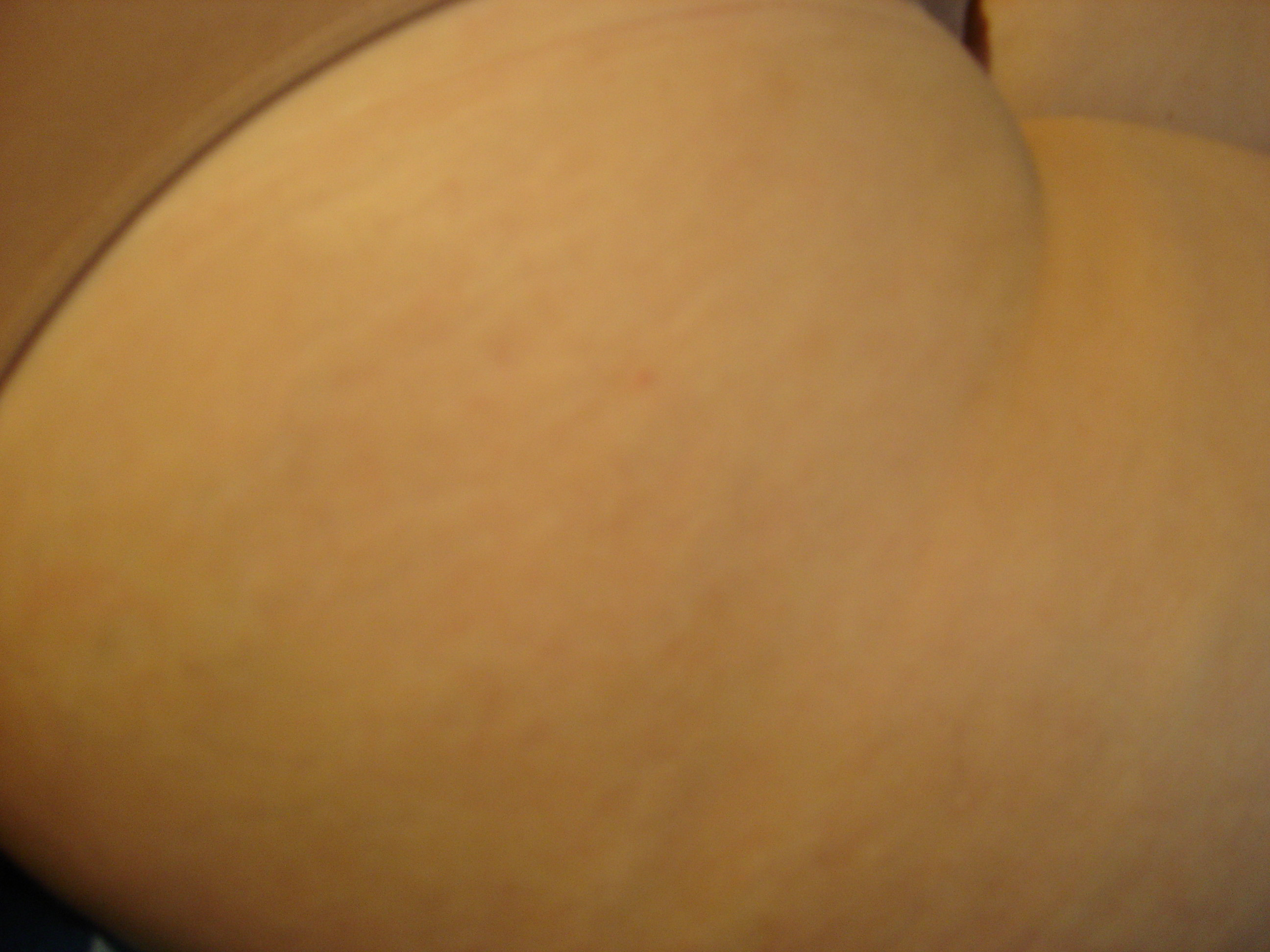


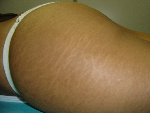
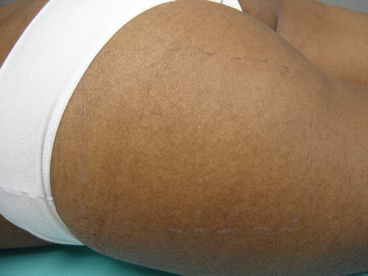
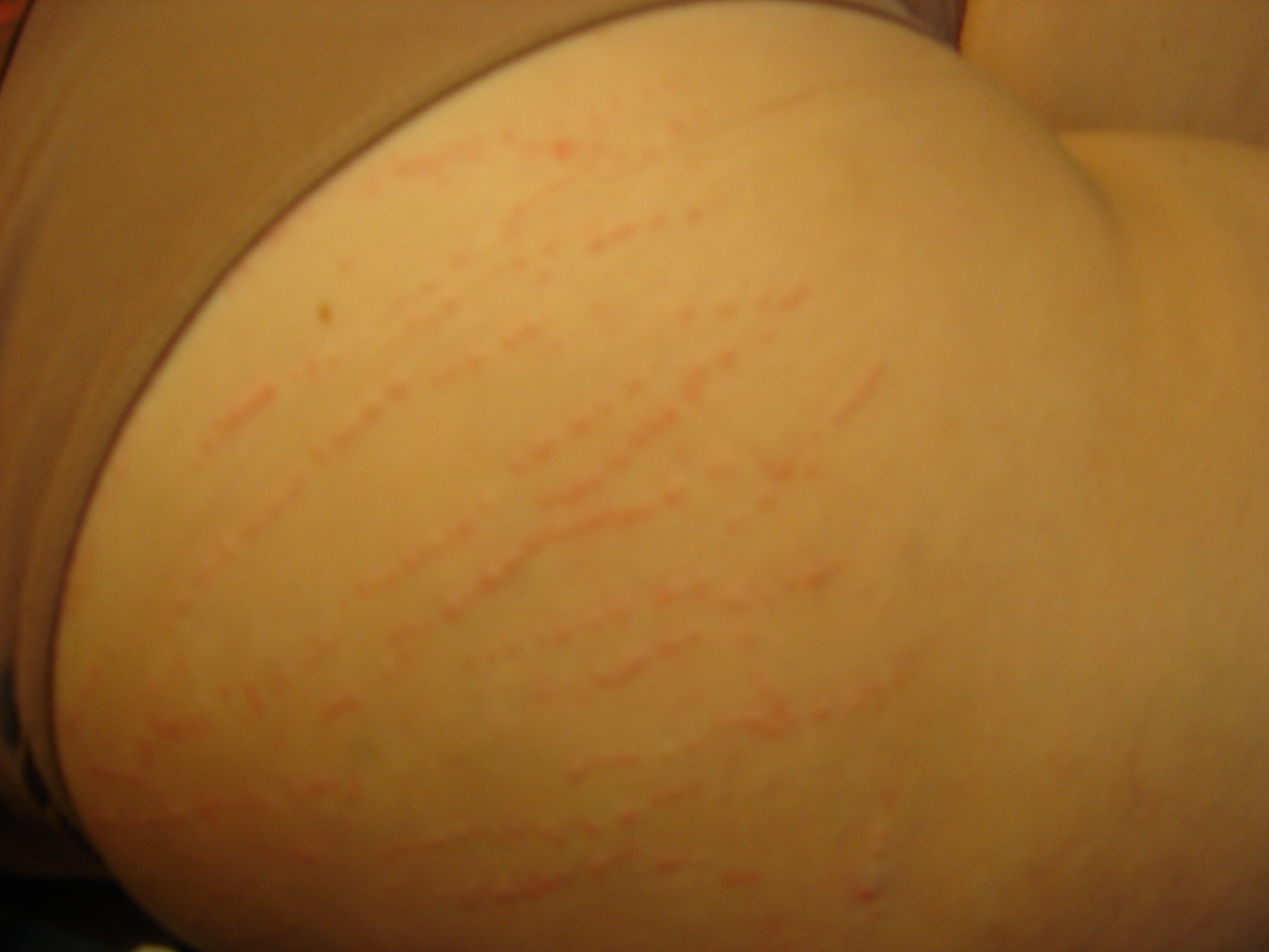
Figure 1. Striae distensae on the buttocks. A) Before treatment. B) Local inflammation immediately after punctures.
Figure 2. Striae alba on buttocks before and after 10-week treatment. A) Appearance before treatment. B) 2 months after last treatment session.
Figure 3. Plasma concentrations of CRP, TNF-α and NO. Values are expressed as mean ± S.E.M.
Figure 4. Plasma concentrations of total and HDL cholesterol. Values are expressed as mean concentration ± S.E.M. Statistics from Bonferroni post hoc test: (a) P<0.05 vs time 0 and 1; (b) P<0.05 vs time 0.
Figure 5. Plasma concentration of TBARS and oxidized LDL-C. Values are expressed as mean ± S.E.M. Statistics from Bonferroni post hoc test: * P<0.01 vs time 0 and 1 for TBARS and P< 0.05 vs time 0 for oxidized LDL-C.
Figure 6. Serum concentrations of CAT and uric acid. Values are expressed as mean concentration ± S.E.M.
3. CONSIDERAÇÕES FINAIS
A estria é um dos problemas estéticos mais comuns encontrados em ambos
os sexos e em todas as idades (2). Apesar de não trazerem nenhum malefício físico,
são esteticamente desagradáveis. Estranhamente, estudos sobre o tratamento dessa
afecção são escassos (9). As opções terapêuticas são muitas, porém, pouco se sabe
sobre os efeitos fisiológicos de cada técnica (10-15). Além disso, a maioria dos
tratamentos possui resultados pouco satisfatórios em estrias albas (9,10).
Pela primeira vez foram investigados os efeitos fisiológicos do tratamento
de estrias com a técnica de galvanopuntura. O presente trabalho mostrou a eficácia da
técnica no tratamento de estrias albas. Melhoras na coloração e aspecto da pele
estriada foram vistas em todas as participantes da pesquisa. Das trinta e duas
participantes, cinco tiveram suas estrias eliminadas por completo antes do término do
tratamento. Provavelmente, a eficácia do método esteja relacionada com a indução da
inflamação aguda e supostamente a ativação dos fibroblastos, com conseqüente
aumento na síntese de colágeno (17).
Devido ao processo inflamatório localizado produzido pela técnica de
galvanopuntura, era esperado que as pacientes tratadas apresentassem algum efeito
inflamatório sistêmico. Contudo, os resultados obtidos neste estudo demonstraram que
nenhum efeito inflamatório sistêmico, tanto agudo quanto crônico, ocorreu durante o
tratamento. Os níveis de dois marcadores inflamatórios, proteína C-reativa (PCR) e
fator de necrose tumoral-α (TNF-α) não mostraram nenhuma alteração significativa
dentro dos tempos analisados. A concentração de NO, outro mediador inflamatório,
também não apresentou alteração significativa, apesar de mostrar uma tendência à
queda logo no início do tratamento.
Segundo Dohy e col. (19), a inflamação tem uma forte relação com o
estresse oxidativo e o perfil lipídico. Com base nessa informação, acreditava-se que o
perfil lipídico dessas pacientes poderia se alterar devido ao aumento do estresse
oxidativo induzido pelo tratamento. Dessa forma, foram avaliados os níveis de
concentração plasmática de colesterol total, lipoproteínas de alta densidade (HDL) e
oxidação das lipoproteínas de baixa densidade (LDL). Os resultados mostraram-se
favoráveis à melhora do perfil lipídico: a concentração de colesterol total não se alterou,
enquanto que as HDL aumentaram e a oxidação das LDL diminuiu. Juntamente com
esses dados, encontramos queda nos níveis das substâncias reativas ao ácido
tiobarbitúrico (TBARS), analisadas como índice de lipoperoxidação.
Para tentar explicar a queda nos níveis dos marcadores de estresse
oxidativo, avaliamos as concentrações de dois importantes antioxidantes: catalase
(CAT) e ácido úrico. A CAT, por estar presente nos peroxissomos, é uma das primeiras
enzimas acionadas no processo de defesa (20). Enquanto que o ácido úrico é o
antioxidante não-enzimático responsável por 50% da capacidade antioxidante no
sangue (20). Entretanto, os níveis séricos desses compostos se mostraram inalterados.
Assim, é necessário buscar outros marcadores que possam estar atuando contra o
Os dados apresentados indicam para a possibilidade da técnica de
galvanopuntura ter um efeito antioxidante e antidislipidêmico. Portanto, o tratamento de
estrias albas através da galvanopuntura demonstra ser eficaz e benéfico para as
pacientes que a utilizam.
4. REFERÊNCIAS BIBLIOGRÁFICAS
Zheng P, Lavker RM, Kligman AM. Anatomy of striae. Br J Dermatol 1985;112:185-93.
Nigam PK. Striae cutis distensae. Int J Dermatol 1989;28:426-8.
Shuster, S. The cause of striae distensae. Acta Derm Venereol 1979;59:161-9 (suppl.85).
Atwal GSS, Manku LK, Griffiths CEM, Polson DW. Striae gravidarum in primiparae. Br J Dermatol 2006;155:965-9.
Cho S, Park ES, Lee DH, Li K, Chung JH. Clinical features and risk factors for striae distensae in Korean adolescents. J Eur Acad Dermatol Venereol 2006;20:1108-13.
Hermanns JF, Piérard GE. High-resolution epiluminescence colorimetry of striae distensae. J Eur Acad Dermatol Venereol 2006;20:282-7.
Davison TE, McCabe MP. Relationships between men's and women's body image and their psychological, social, and sexual functioning. Sex Roles 2005;52(7/8):463-75.
Gingras J, Fitzpatrick J, McCargar L. Body image of chronic dieters: lowered appearance evaluation and body satisfaction. J Am Diet Assoc 2004;104:1589-92.
Jiménez GP, Flores F, Berman B, Gunja-Smith Z. Treatment of striae rubra and striae alba with the 585-nm pulsed-dye laser. Dermatol Surg 2003;29:362-5.
10. Nouri K, Romagosa R, Chartier T, Bowes L, Spencer JM. Comparison of the
585 nm pulse dye laser and the short-pulsed CO2 laser in the treatment of striae distensae in skin types IV and VI. Dermatol Surg 1999;25(5):368-70.
11. Alexiades-Armenakas MR, Bernstein LJ, Friedman PM, Geronemus RG. The
safety and efficacy of the 308-nm excimer laser for pigment correction of hypopigmented scars and striae alba. Arch Dermatol 2004;140:955-60.
12. Ash K, Lord J, Zukowski M, McDaniel DH. Comparison of topical therapy for
striae alba (20% glycolic acid/ 0,05% tretinoin versus 20% glycolic acid/ 10% L-ascorbic acid). Dermatol Surg 1998;24:849-56.
13. Kang S, Kim KJ, Griffiths CEM, Wong T-Y, Talwar HS, Fisher GJ, et al. Topical
tretinoin (retinoic acid) improves early stretch marks. Arch Dermat 1996;132(5):519-26.
14. Spencer JM. Microdermabrasion. Am J Clin Dermatol 2005;6(2):89-92.
15. Guirro ECO, Guirro RRJ. Estrias. In: Fisioterapia dermato-funcional:
fundamentos, recursos, patologias, 3ª ed. Barueri,SP:Manole. 2004; 391-412.
16. Van Leeuwen MA, Van Rijswijk MH. Acute-phase proteins in the monitoring of
inflammatory disorders. Baillieres Clin Rheumatol 1994;8(3):531-52.
17. Weiss DS, Kirsner R, Eaglstein WH. Electrical stimulation and wound healing.
Arch Dermatol 1990;126:222-5.
18. Gabay C, Kushner I. Acute-phase proteins and other systemic responses to
inflammation. N Engl J Med 1999;340(6):448-54.
19. Dohy Y, Takase H, Sato K, Ueda R. Association among C-reactive protein,
oxidative stress, and traditional risk factors in healthy Japanese subjects. Int J Cardiol 2007; 115:63-6.
20. Somogyi A, Rosta C, Pusztai P et al. Antioxidant measurements. Physiol Meas
21. Benzie IFF. Evolution of antioxidant defense mechanisms. Eur J Nutr
22. Stuart-Smith K. Demystified nitric oxide. J Clin Pathol: Mol Pathol
5. ANEXO 1: DOCUMENTO DE CONFIRMAÇÃO DE SUBMISSÃO
Source: http://tede.pucrs.br/tde_busca/processaArquivo.php?codArquivo=943
Que mediante la Resolución No. 852002 del 19 de junio de 2002, el Consejo Arancelario y Aduanero Que conforme los artículos 38, 39 y 55 del Protocolo al Tratado General de Integración Económica Centroamericana Protocolo de Guatemala, modificado por la Enmienda del 27 de febrero de 2002, el Consejo de Ministros de Integración Económica (COMIECO), tiene bajo su competencia los asuntos de la Integración Económica Centroamericana y como tal, le corresponde aprobar los actos administrativos del Subsistema Económico;
Report of the Independent Expert in the Matter of the Public Utilities Commission Initial Decision In the 2008 Annual Review Proceeding for Belize Electricity Limited Jonathan A. Lesser, Ph.D.* Initial Draft: 6 June 2008 Final Report: 11 June 2008 * Jonathan A. Lesser, Ph.D., is a Partner with Bates White, LLC, an economic and litigation consulting firm headquartered in Washington, DC. However, the views, findings, conclusions, and recommendations expressed herein are those of Dr. Lesser alone, and do not necessarily reflect the views of his colleagues or of Bates White, LLC.


