Need help?

800-5315-2751 Hours: 8am-5pm PST M-Th;  8am-4pm PST Fri
Medicine Lakex
medicinelakex1.com
/a/apmfc.org.br1.html
 

00405 1.13

Original article 1 Chronic inhibition of NO synthesis per se promotes structural intimal remodeling of the rat aorta Marcos A. Rossi and Massimo Colombini-Netto Objective We characterized, using histomorphometry and thickened (60% increase in comparison with that of transmission and scanning electron microscopy, the controls and 65% thinner as compared with L-NAME- intimal remodeling of the thoracic aorta of treated rats). In the same way, the mean intima:media ratio normocholesterolemic young rats chronically-treated with of rats concomitantly treated with L-NAME and captopril L-NAME and examined the question whether these was moderately increased (45% more) as compared with changes were caused by the lack of NO per se or by the controls and signi®cantly lower in comparison with rats hypertension which L-NAME administration induces.
administered L-NAME alone (36% less).
Methods Male Wistar rats were divided randomly into Conclusions Chronic inhibition of NO synthesis per se three sets: control group, standard diet/L-NAME-treated promotes structural intimal remodeling of the rat aorta, group, and standard diet/L-NAME captopril-treated which is potentiated by L-NAME-induced hypertension.
Most important, the present ®ndings favor the idea thatblockade of NO synthesis by causing intimal remodeling Results The treatment of rats with L-NAME for 4 weeks might be a primary cause, as individual biologic resulted in increased blood pressure (by 32% at the end of phenomenon, in the development of an atherosclerotic the treatment) as compared with the control value and plaque. J Hypertens 19:1±13 & 2001 Lippincott Williams & intimal remodeling comprising a continuous layer of enlarged endothelial cells with irregular nuclear andcytoplasmic contours, lying over a thickened layer of Journal of Hypertension 2001, 19:1±13 ®brocollagenous support tissue focally expanded withlymphomononuclear cells and mainly diffuse foci of Keywords: atherosclerosis, endothelium, hypertension, intima, L-NAME smooth muscle cells. In addition, the NO synthase hypertension, nitric oxide, NO, NO synthase, remodeling inhibition caused a marked thickened tunica intima (150% Department of Pathology, Faculty of Medicine of RibeiraÄo Preto, University of SaÄo thicker than the control value) and a signi®cantly Paulo, RibeiraÄo Preto, SP, Brazil augmented intima:media ratio (126% higher than the Sponsorship: Supported by a grant from the FundacËaÄo de Amparo aÁ Pesquisa control value). On the other hand, captopril prevented do Estado de SaÄo Paulo (FAPESP 99/08574±7).
hypertension in rats simultaneously treated with L-NAME Correspondence and requests for reprints to: Marcos A. Rossi, Professor of as compared with controls, and induced intimal Pathology, Department of Pathology, Faculty of Medicine of RibeiraÄo Preto, University of SaÄo Paulo, 14049±900 RibeiraÄo Preto, SP, Brazil.
remodeling comprising the same qualitative changes as Tel: ‡55 15 602 3130; fax: ‡55 15 602 3130; e-mail: [email protected] those observed in L-NAME-treated rats. The tunica intima Received 7 September 2000 Revised 20 April 2001 of L-NAME captopril-treated rats was moderately Accepted 2 May 2001 hypertension, profoundly affects endothelial function Atherosclerosis is now believed to be an in¯ammatory that manifests as diminished release of nitric oxide into process driven by in¯ammatory cells, in particular the arterial wall either because impaired local produc- macrophages, in association with subendothelial (inti- tion or excessive oxidative degradation Impor- mal) deposition of lipids in the artery wall The tantly, however, it remains unclear whether this characteristic lesion of atherosclerosis represents the endothelial dysfunction manifestation is a cause or a result of cycles of accumulation of monocyte-derived consequence of lipid accumulation since fatty streaks macrophages and migration and proliferation of smooth are also present from a young age.
muscle cells that occur principally in the tunica intima of large and medium-sized elastic and muscular arteries.
Recent studies have shown that chronic administration Endothelial dysfunction is an early feature in athero- of L-arginine analogues such as Nù-nitro-L-arginine sclerosis that manifests as impaired local nitric oxide methyl ester (L-NAME) to rats induces a dose depen- (NO) production and increased expression of adhesion dent systemic arterial hypertension, decreased intra- molecules It has been clear that risk factors for cellular cGMP levels (the second messenger of NO) in atherosclerosis, such as hypercholesterolemia and vascular smooth muscle, and in¯ammatory phenotypic 0263-6352 & 2001 Lippincott Williams & Wilkins Article number = 00405 2 Journal of Hypertension 2001, Vol 19 No 8 changes in the coronary vascular wall Since L-NAME ‡ captopril groups at the beginning (consid- NO-donor compounds have been shown to inhibit ered as a uni®ed 0 week time point, n ˆ 35) and at the vascular smooth muscle cell proliferation, protein synth- end of the experiment (4 week time points relatives to esis, and production of extracellular matrix in culture control, n ˆ 7, L-NAME-treated, n ˆ 11, and L- and to restrict proliferation and mitogenesis of NAME ‡ captopril-treated, n ˆ 9, groups) were used to cultured endothelial cells the present study was measure the concentration of total and HDL cholester- designed to determine the effect of prolonged NO ol by a spectrophotometric method using a commer- synthase inhibition with L-NAME on the intima of the cially available kit (Cobas Mira, Roche). Serum proteins aorta in rats. We characterized, using histomorphometry and albumin levels were estimated at the end of the and transmission and scanning electron microscopy, the experimental period by means of the biuret reaction.
intimal remodeling of the thoracic aorta of normocho- lesterolemic young rats and examined the question Mean arterial blood pressures were measured once a whether these changes were caused by the lack of NO week by a tail cuff method without anesthesia. The per se or by the hypertension which L-NAME adminis- mean of three measurements was considered to be the tration induces. This way, the main objective was to mean for the week. A tail cuff was placed over the tail detail the reduction of NO availability, as individual with the systolic and diastolic blood pressure being biological phenomenon, on early aortic intimal remo- measured on a NARCO physiograph (E & M Instru- ments Co., Inc, Houston, Texas, USA). Measurements were made after the rat had been resting for a few minutes in a hot box.
Male Wistar albino rats, weighing an average of 87 g, were obtained from the breeding colony at the Faculty After 4 weeks, the rats were killed in light ether of Medicine of RibeiraÄo Preto. The animals were anesthesia by exsanguination from the abdominal aorta, housed individually in cages constructed of polypropy- between 0900 and 1100 h. The thoracic cavity was lene with stainless steel mesh tops washed twice opened exposing the still beating heart. The hearts weekly. The rats were fed solid laboratory rat food in were rapidly removed, rinsed in ice-cold 0.9% NaCl stainless steel feeding dishes and liquid in Richter solution (saline), blotted and weighed. The heart graduated drinking tubes. Their liquid intake was weight index was calculated by dividing the heart recorded thrice weekly and their solid food consump- weight to body weight (heart ratio). The hearts were tion was recorded twice weekly. The approximate ®xed as a whole in neutral 10% formalin for histological composition of the commercial standard diet (g/100 g) study. Both ventricles of each heart were isolated and was: protein 21.0, fat 3.5, carbohydrate 60.0, salts 5.3, cut into three fragments by two coronal sections at vitamin mixture 2.0, and remainder ash (the data were equal intervals. Each block was serially cut at 6 ìm in provided by the supplier). This diet provided approxi- the same direction, and sections were stained with mately 3.5 kcal/g. The animals were divided randomly hematoxylin and eosin and picrosirius red technique.
into three sets: (1) a control group, drinking untreated The quantitative examination of the left myocardium water and receiving a dose of placebo (1 ml of sterile was carried out on medium power light-microscopic saline solution administered intra-peritoneally); (2) a ®eld ( 3 320): 100-indexed square graticle (Carl Zeiss, standard diet/L-NAME-treated group, drinking water Jena, Germany) was used to estimate the volume containing 45 mg/100 ml of Nù-nitro-L-arginine methyl fraction (% of ®brosis in picrosirius red-stained sections.
ester, L-NAME and a daily dose of placebo (1 ml of Fields (n ˆ 48) of subepicardial and midmyocardial sterile saline solution administered intra-peritoneally); zones were analyzed for each heart (seven hearts from (3) and a standard diet/L-NAME ‡ captopril-treated control group, 11 hearts from L-NAME group and six group, drinking water containing 45 mg/100 ml of L- hearts from L-NAME ‡ captopril group).
NAME and administered intra-peritoneally a daily dose of captopril, an angiotensin converting enzyme inhibi- The aortas (n ˆ 6) were rinsed at a mild pressure- tor, at 40 mg/kg of body weight. The animals were perfusion of 100 mmHg with phosphate buffered saline weighed twice weekly. Captopril was freshly prepared (PBS) through the ascending aorta followed by perfu- by dissolving the drug powder in 1 ml of sterile saline sion ®xation with 2.5% glutaraldehyde in cacodylate solution and administered daily via intra-peritoneal buffer (pH 7.3) for 3 min. The thoracic aortas were then injection. The administration of captopril began 3 days immediately harvested (n ˆ 6 in each group) and the before L-NAME administration in the drinking water.
adventitial tissue was removed. The aortic tubes were The dosage used was based on pilot experiments and transversally cut into 5±6, 2 mm-long fragments (sam- was found to be effective for blood pressure control, as pling from the upper to the lower part of the thoracic previously reported aorta) and ®xed in the same ®xative for 2 h. The samples were rinsed in cacodylate buffer, post®xed in The serum of rats from control, L-NAME-treated, and 1% osmium tetroxide buffered in cacodylate for 2 h, NO synthesis inhibition per se promotes aortic intimal remodeling Rossi and Colombini-Netto 3 dehydrated in ascending concentrations of acetone, and nia, USA) for an IBM PC computer. For analysis, one- subsequently embedded in Araldite1. Morphometric way analysis of variance (ANOVA) and the Bonferroni analysis was performed using videomicroscopy with the test (to correct for multiple comparisons) were used.
Kontron Image System KS-100 software (Kontron Elek- One value for each case entered into the analysis. A tronik GmbH, Germany) in conjunction with a Zeiss level of signi®cance of 5% was chosen to denote the microscopy, video camera, and an on-line computer.
difference between group means. Unless speci®ed, data Cross sections (n ˆ 5±6) of good technical quality, are presented as mean  standard error.
exactly perpendicular to the long axis of the aorta from each vascular segment, were chosen for morphometric evaluation. The absolute thickness of the intima, as Rats treated with L-NAME or L-NAME ‡ captopril de®ned as the endothelium and the subendothelial remained in good health, with no signs of nutri- layer to the inner edge of the internal elastic lamina tional de®ciencies. However, the L-NAME- and L- (IEL), the absolute thickness of the media, as de®ned NAME ‡ captopril-treated rats gained less weight by the tissue between the IEL and the external elastic (6.65 and 6.27 g/day per rat, respectively) as com- lamina (EEL), and the cross sectional area of the aorta pared with controls (8.07 g/day per rat) over the 4- lumen were measured. The number of measurements week period. The mean ®nal body weight of control to estimate the intima and media thickness was 100 animals was 314.10 g, respectively 13% and 16.5% around the vessel circumference for each aorta from higher than the ®nal body weight of L-NAME- each rat. This number was chosen arbitrarily. The use L-NAME ‡ captopril-treated of thin plastic sections stained with toluidine blue allowed adequate resolution of structural details. Meas- urements were made by a skilled observer blinded to The animals in the present study received L-NAME the treatment groups. The intima:media ratio was dissolved in the drinking water, 45 mg/100 ml. Mean calculated by dividing the intima thickness to media water intake in the control, L-NAME- and L-NAME - thickness and multiplying per 100. Intima and media ‡ captopril groups decreased from 19.27, 19.96, and thickness was determined with the use of an overall 21.77 ml/day per 100 g of body weight at the beginning microscope magni®cation of 3 400 whereas the cross- of the experiment to 11.63, 15.02, and 13.51 ml/day per sectional area was determined at 3 100. Findings were 100 g at the end, respectively. The average intake of averaged for each group. Ultrathin sections were ob- drinking water did not differ signi®cantly in both L- tained from selected areas with a diamond knife in a NAME- (18.16 ml/day per 100 g) and L-NAME ‡ cap- Sorvall MT-5000 ultramicrotome (Du Pont Co., Wil- topril-treated animals (19.04 ml/day per 100 g while the mington, Delaware, USA) stained with uranyl acetate average intake of drinking water in controls (15.98 ml/ and lead citrate, and examined in a Zeiss EM 109 day per 100 g) was slightly smaller. The mean intake of transmission electron microscope (Carl Zeiss, Oberko- L-NAME during the experimental period decreased chen, Germany) at 80 kV.
from 8.98 and 9.80 mg/day per 100 g of body weight at the beginning of the experiment to 6.75 and 6.07 mg/ The thoracic aortas of four animals from each group day per 100 g at the end in L-NAME- and L-NAME - were immediately harvested and cut into anterior and ‡ captopril-treated rats, respectively. The average in- posterior halves. Fragments 5±6 mm long were ®xed by takes of L-NAME in both L-NAME-tested rats were immersion in phosphate buffer 2.5% glutaraldehyde similar (8.17 ml/day per 100 g in L-NAME-treated and (pH 7.3) for 2 h, post®xed in osmium tetroxide in 8.57 ml/day per 100 g in L-NAME ‡ captopril-treated phosphate buffer for 2 h, dehydrated in ascending rats). In contrast, average consumption of solid food in concentrations of ethanol, and dried in liquid carbon control rats (19.54 g/day per rat) was higher than the dioxide by the critical point method. To improve our average consumption in L-NAME- (17.37 g/day per rat) visualization of the endothelial cells, the specimens and in L-NAME ‡ captopril-treated rats (16.32 g/day already ®xed were put into 1% HCl for 30 s with gentle shaking. The dried specimens were then glued on aluminum stubs with silver paste, sputter-coated with At the ®rst day preceding the beginning of intra- gold, and examined in a Zeiss 940A scanning electron peritoneal injections of captopril and placebo, the microscope at 15 kV. The segments were ®xed under plasma concentration of total and HDL cholesterol atmospheric pressure rather than physiological pressure (0 week time point) was 59.80  4.54 mg/100 ml, and to prevent eventual artifacts due to overstretching. It is 39.27  2.34 mg/100 mg, respectively. These values felt that qualitative assessment of this data is justi®ed, were closely similar to those found at the 4 week time as all segments were prepared under similar conditions.
point of control (60.33  5.32 mg/100 ml of cholesterol and 43.63  2.60 mg/100 ml of HDL cholesterol), of L- Data were analyzed using a GraphPad Prism statistic NAME-treated rats (58.14  3.72 mg/100 ml of choles- program (GraphPad Software Inc., San Diego, Califor- terol and 43.51  1.87 mg/100 ml of HDL cholesterol),


4 Journal of Hypertension 2001, Vol 19 No 8 and L-NAME ‡ captopril-treated rats (62.63  4.73 mg/ cross-sectional luminal area of aortas from L-NAME- 100 ml of cholesterol and 41.05  3.11 mg/100 ml of and L-NAME ‡ captopril-treated rats was 1.34  HDL cholesterol. The total plasma proteins and albu- 0.04 mm2 and 1.30  0.03 mm2, respectively, quite si- min levels of L-NAME-treated rats (5.76  0.41 g/ milar to the mean cross-sectional area of control aortas, 100 ml and 3.42  0.13 g/100 ml, respectively) and L- 1.27  0.04 mm2.
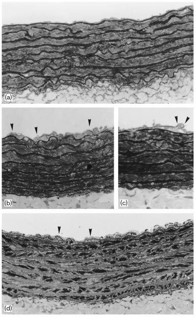
NAME ‡ captopril-treated rats (6.33  0.42 g/100 ml and 3.57  0.12 g/100 ml, respectively) were closely The light microscopic study of plastic-embedded aortas similar to those of controls (5.55  0.54 g/100 ml of total could demonstrate striking intimal changes in plasma proteins and 3.53  0.18 g/100 ml of albumin).
and L-NAME ‡ captopril-treated groups as compared with control group (Fig. The Treatment with L-NAME only, increased moderately changes in the L-NAME-treated groups were character- the mean arterial blood pressure, by approximately 32% ized by enlarged endothelial cells lying over a thick in comparison with that of untreated controls.s and extracellular toluidine blue stained support tissue and 3.53  0.18 g/100 ml of albumin).
Treatment with L-NAME only, increased moderately the mean arterial blood pressure, by approximately 32% in comparison with that of untreated controls. The concomitant treatment with L-NAME and captopril decreased the mean arterial blood pressure by approxi- mately 4% versus control animals. The hypertensive response was time-dependent reaching a maximum at the week 4. Captopril was able to inhibit the develop- ment of high blood pressure since the ®rst week of treatment with L-NAME.
The hearts from rats of groups control, L-NAME, andL-NAME ‡ captopril were similar on gross examina- tion, and no differences were found in their weights expressed in g/100 g body weight: 3.35  0.10 and 3.41  0.11 and 3.42  0.07, respectively. Since the body weight of animals is an important source of variability of organ weight, it seems obvious that the organ weight should be corrected for differences in body weights. Body and heart weight data on a large group of normal male rats in the body weight range of 50 g and 450 g were collected, and expressed as a weight curve of heart relative to body weight By using this method, it was possible to compare the wet heart weight of rats from both groups to wet heart weight of equal body weight predicted controls. The wet heart weight of L-NAME-treated (924.82  L-NAME ‡ captopril-treated 36.13 mg), and control (1050.08  29.63 mg) were not different from the predicted wet heart weight of equal body weights controls, 920, 882, and 1005 mg, respec- tively. The analysis of picrosirius red-stained sections revealed mild myocardial ®brosis manifested by dis- crete increase in amount of pericellular collagen (en- domysial matrix) and mild perivascular ®brosis. The volume fraction of ®brosis of the controls was 2.56  0.30% whereas the volume fraction of L-NAME Light microscopy. Representative views of aorta walls from (a) control, (b,c) L-NAME-and (d) L-NAME ‡ captopril-treated rats. Glutaraldehyde- and L-NAME ‡ captopril hearts increased to 4.07  ®xed, plastic-embedded tissue section stained with toluidine blue. (a) 0.18% and 4.75  0.28% illustrates the delicate intima of control rats. A clearly expanded intima can be seen in b, c and d. Clusters of smooth muscle cells (b and d arrowheads) and mononuclear cells (c arrowheads) within the intima Grossly, the thoracic aortas from both experimental can be seen. (Mag. 3 410).
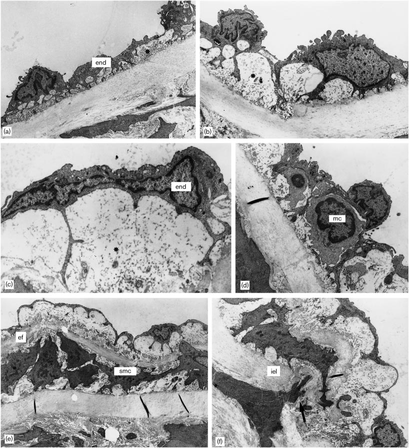
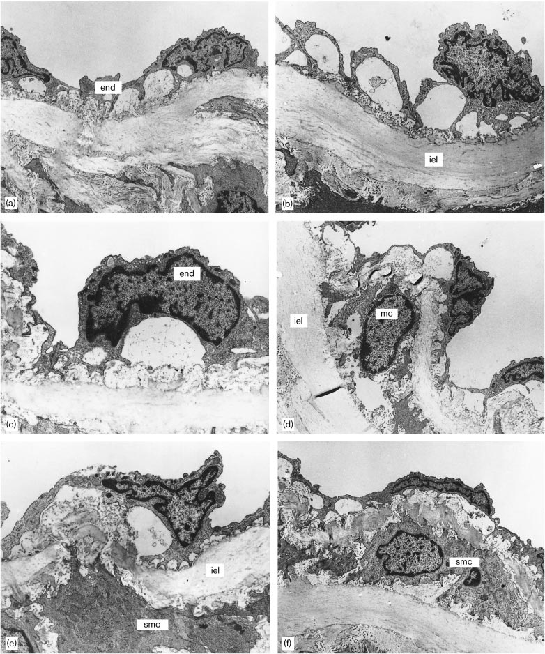
groups appeared similar to those of controls. The mean NO synthesis inhibition per se promotes aortic intimal remodeling Rossi and Colombini-Netto 5 foci of intimal thickening. This was composed of appeared enlarged with irregular nuclear and cytoplas- mononuclear cells or multilayered cushions of smooth mic contours resting on a basement membrane-like muscle cells in contrast to the delicate structure of the material. Beneath this there was a thicker layer of intima in the control group. Simultaneous treatment of ®brocollagenous support tissue. Clusters of smooth the rats with L-NAME and captopril resulted in clearly muscle cells appeared within the intima, randomly attenuated qualitatively similar intimal changes. This arranged, and surrounded by basement membrane-like could be clearly demonstrated by evaluating the tunica material, collagen and young elastic ®bers. Migration of intima and tunica media thickness and the intima: smooth muscle cells from the media into the intima media ratio by means of light microscopy morphometry.
through the fenestras of the internal elastic lamina Light microscopy sampling in morphometry was pre- could be also seen. Diffuse focal accumulation of ferred because large test areas could be analyzed. The mononuclear cells was detected in the expanded in- use of thin plastic allowed adequate resolution of timal layer. The media remained intact and the medial structural details. The histomorphometric analysis smooth muscle cells appeared unaltered and retained showed that the mean intima thickness of the aorta their original orientation to the vessel from rats receiving L-NAME was 4.32  0.33 ìm, 150% higher than the value 1.73  0.11 ìm determined for In the L-NAME ‡ captopril-treated rats the intima was the controls, and 54% higher than the value also thickened as compared with controls. Ultrastructur- 2.81  0.17 ìm determined for the rats concomitantly al changes were qualitatively the same as those found given L-NAME and captopril. In the same way, the in the L-NAME-treated animals, although less pro- intima:media ratio in L-NAME-treated rats (4.22  nounced as clearly suggested by the histomorphometric 0.38) was 126% increased as compared with control rats (1.87  0.14) or 56% increased as compared with the mean ratio for rats administered L-NAME and captopril The scanning electron microscopic evaluation revealed (2.71  0.19). On the other hand, the mean that the endothelial surface was lightly wavy. The media thickness of the thoracic aorta from L-NAME- endothelial cells throughout the aorta of control animals treated (103.00  3.99 ìm), L-NAME ‡ captopril-trea- were elongated in shape with a major axis parallel to ted (104.3  4.92 ìm), and control rats (92,80  the direction of the blood ¯ow. Marginal folds marked 1.27 ìm) were not statistically different.
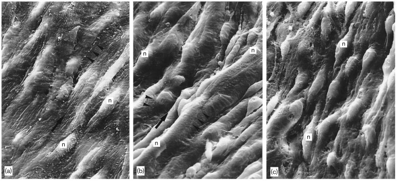
the margins. The luminal surface had microvilli spread out, mainly clustered over the nuclear hillock. The The body weight (initial and ®nal), growth rate, heart endothelial surface of the aorta of L-NAME-treated rats weight, heart ratio, and mean arterial blood pressure was highly irregular with a striking nodular pattern with (initial and ®nal) from the three groups are tabulated in deep grooves between the nodules while the endothe- The average daily intake of water and L- lial surface of the aorta of rats concomitantly treated NAME, solid food consumption, and the mean plasma with L-NAME and captopril was moderately irregular concentration of total and HDL cholesterol, and the with discrete nodular pattern. In both groups, the total plasma proteins and albumin levels for all three endothelial cells were ill-de®ned, with less evident groups are given in s the values margins due to the paucity of marginal folds (Fig. of tunica intima and tunica media thickness, intima:me- dia ratio and cross sectional luminal area of the aorta from control, L-NAME-treated, and L-NAME ‡ capto- In the present study, we show that inhibition of NO pril-treated groups. This can be clearly seen when the synthesis by chronic administration of L-NAME into frequency distribution of intima and media thickness rats induces hypertension and intimal remodeling in and intima: media ratios in each group were plotted the absence of elevated plasma cholesterol levels. In addition, and importantly, we found that animals made normotensive by simultaneous L-NAME and captopril The transmission electron microscopic appearance of treatment had reduced, although qualitatively un- the intima of control aortas did not differ from that changed, intimal remodeling.
reported in the literature. The intima was composed of a continuous layer of endothelial cells sitting on a basal In our studies, Nù-nitro-L-arginine methyl ester (L- lamina and a very thin layer of ®brocollagenous support NAME) was given in the drinking water containing tissue. This is contiguous with the media, composed of 45 mg/100 ml, resulting in a mean daily intake of smooth muscle cells reinforced by organized layers of approximately 8 and 8.5 mg/100 g of body weight for L- elastic ®bers forming elastic laminas and collagen ®bers.
NAME- and L-NAME ‡ captopril-treated rats, respec- Just below the intima there is a broad continuous layer tively. However, intake of L-NAME decreased from of elastic tissue interrupted by fenestras, the internal the beginning of the treatment period to the end, as elastic lamina (Fig. In the L-NAME-treated rats the previously reported It has been demonstrated that intima was diffusely expanded. The endothelial cells endothelial NO synthesis is inhibited by L-NAME 6 Journal of Hypertension 2001, Vol 19 No 8 Body weights (initial and ®nal), growth rates, heart weights, heart ratios, and mean arterial blood pressure (initial and ®nal) from control, L-NAME-treated, and L-NAME captopril-treated rats Heart weight (mg) Mean blood pressure 88.14  1.30 314.10  8.48 77.82  7.48 107.5  7.46 L-NAME-treated (n ˆ 11) 87.14  1.02 273.40  8.25 79.69  8.46 141.7  7.46 L-NAME ‡ captopril-treated 86.05  1.22 261.16  6.11 80.20  9.21 101.4  6.43 NAME ‡ captoprilL-NAME 3 L-NAME ‡ captopril Values are mean  standard error. Measured versus predicted. Mean  standard deviation. NS, not signi®cant.
Average daily intake of water and L-NAME, solid food consumption, and mean plasma concentration of total and HDL cholesterol, and total plasma proteins and albumin levels from control, L-NAME-treated, and L-NAME captopril-treated rats.
Water intake L-NAME intake L-NAME-treated (n ˆ 11) L-NAME ‡ captopril-treated (n ˆ 9) Values are mean  standard error.
Tunica intima and media thickness, intima:media ratio, and cross sectional area of the aorta from control, L-NAME-treated, and L-NAME captopril-treated rats Intima:media ratio L-NAME ‡ captopril (n ˆ 6) P Control 3 L-NAME ‡ captopril L-NAME 3 L-NAME ‡ captopril Values are mean  standard error of the mean. NS, not signi®cant administration. Treatment of adult rats with a daily L-arginine normalized the L-NAME-induced decrease dose of 4 mg/day per 100 g of body weight for 4 weeks, in NO production alone or with captopril has been shown to result in a signi®cant attenuation of NO synthase activity in L-NAME-treated rats as compared with controls experi- the heart (69% less), aorta (26% less), brain (73% less), enced mild retardation of body weight gain. It could be and kidney (30% less). L-NAME administered in the argued that the effect of NO synthase inhibition could drinking water (50 mg/100 ml) to rats markedly reduced be related to insuf®cient nutrition. This is very unlikely the levels of NO metabolites in serum and urine while since the serum albumin levels of L-NAME-treated rats increasing blood pressure. The simultaneous adminis- were closely similar to those of control rats and tration of an angiotensin converting enzyme inhibitor hypoalbuminemia is an invariable ®nding in developed completely normalized the blood pressure without protein-calorie under nutrition Similarly, rats affecting the decreased NO production Further- chronically treated with 20±80 mg/100 ml in the drink- more, the NO production from the aortas of rats treated ing water of L-NAME or rabbits chronically treated with L-NAME (10 mg/day per 100 g of body weight) with 80 mg/100 ml of L-NAME in the drinking water, was markedly less than in control group aortas, whereas did not shown alterations of serum levels of albumin


NO synthesis inhibition per se promotes aortic intimal remodeling Rossi and Colombini-Netto 7 Frequency distribution ( Intima : media ratio Frequency distribution ( Intima thickness (µm) Frequency distribution ( Media thickness (µm) Frequency distribution of intima:media ratio and tunica intima and media thickness in L-NAME-treated (ÐÐÐÐ), L-NAME ‡ captopril- treated (± ± ±), and control (´ ´ ´ ´ ´ ´) groups.
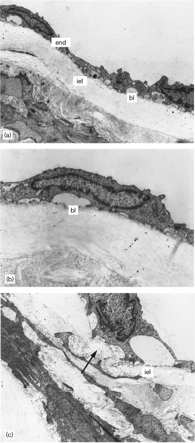
Transmission electron microscopy. Control aorta. (a) The delicate structure of the intima contains ¯attened endothelial cells (end) sitting In addition, the observed daily food consump- on a basal lamina (bl) and a very thin layer of support tissue (); (iel), tion was in accord with the assumed adequate dietary internal elastic lamina. (b) a higher magni®cation of one of the intake of 15 g/day per rat for growing rats or adult rats endothelial cells seen in (a). (c) The intima is delimitated by the internal elastic lamina (iel) interrupted by fenestras (arrow). (Mags: a, 3 4350; at maintenance. The slightly higher average con- b, 3 10450; c, 3 5800.) sumption of solid food in control rats, in comparison


8 Journal of Hypertension 2001, Vol 19 No 8 Transmission electron microscopy. L-NAME-treated aorta. (a,b) The intima is thickened. The endothelial cells (end) are heterogeneous, most of them enlarged with convoluted nuclear and cytoplasmic contours. The endothelial cells are sitting over a thicker layer of support tissue (). (c) a higher magni®cation of an enlarged endothelial cell clearly showing tortuous nuclear and cytoplasmic contours. Focal accumulation of mononuclear cells (mc) could be detected in the expanded intimal layer, as seen in (d). (e) Clusters of smooth muscle cells (smc) appear within the intima, randomly arranged, and surrounded by basement membrane-like material, collagen and young elastic ®bers (ef). Migration of smooth muscle cells from the media into the intima (arrow) through the fenestras of the internal elastic lamina (iel) can be also seen (panel F). (Mags. a, 3 4350; b, 3 6900; c, 3 10450; d, 3 5750; e, 3 3500; f, 3 5750.)


NO synthesis inhibition per se promotes aortic intimal remodeling Rossi and Colombini-Netto 9 Transmission electron microscopy. L-NAME ‡ captopril-treated aorta. The ultrastructural changes are qualitatively similar to those found in the L- NAME-treated animals, although less pronounced. (a,b,c) The tunica intima layer is expanded. The endothelial cells (end) are heterogeneous, most of them enlarged with tortuous nuclear and cytoplasmic contours. A mononuclear cell (mc) is seen in the thickened intimal layer in (d). A smooth muscle cell migrating through the fenestra of the internal elastic lamina is illustrated in panel e and clusters of smooth muscle cells (smc) surrounded by collagen and elastic ®bers can be seen in (f). iel, internal elastic lamina. (Mags. a, 3 5300; b, 3 6500; c, 3 9600; d, 3 5800; e, 3 4850; f,


10 Journal of Hypertension 2001, Vol 19 No 8 Scanning electron microscopy. (a) Control aorta. The endothelial cells are elongated in shape with major axis parallel to the direction of the blood ¯ow (thick arrow). Marginal folds (arrowheads) distinctly mark the margins. The nuclear hillocks can be clearly seen (n). (b) L-NAME-treated aorta.
The endothelial surface is highly irregular with striking nodular pattern with deep grooves between the nodules. The endothelial cells are ill- de®ned with less evident margins due to paucity of marginal folds (arrowheads). The nuclear hillocks are more pronounced in comparison to controls (n). (c) L-NAME ‡ captopril-treated aorta. The endothelial surface is moderately irregular with discrete nodular pattern, although qualitatively similar to the appearance shown in panel (b). (Mag. 3 2050.
with the average consumption in L-NAME-tested rats, terized by areas of dense and replacement ®brosis may be related to the ability of NO regulating appetite consistent with organized myocytolytic necrosis and The mean wet heart weights of rats given either striking perivascular ®brosis The interstitial L-NAME or L-NAME and captopril were signi®cantly myocardial ®brosis would be related to L-NAME-in- decreased in comparison with those of controls while duced vasoconstriction with consequent myocardial the heart ratios of all three groups were not different.
ischemia Previous studies addressing the ques- However, the mean wet heart weights of both L- tion whether the myocardial ®brosis during NO NAME-tested rats and of control animals were not synthase inhibition was caused by the consequent different from the predicted heart weight of equal body hypertension or the lack of NO per se reported con¯ict- weight controls. Similarly, several authors did not ®nd ing results. Concomitant administration of hydralazine any hypertrophy of the whole heart or the left and L-NAME prevented arterial hypertension and ventricle after oral L-NAME treatment for 4± attenuated myocardial ®brosis without affecting cardiac 8 weeks. Negative metabolic effects of L-NAME on hypertrophy and microvascular remodeling induced by protein synthesis in spite of the induced sustained chronic inhibition of NO synthesis Co-treatment pressure overload would mediate this blunted with enalapril (angiotensin converting enzyme inhibi- hypertrophic response. This same mechanism could tor) and L-NAME prevented arterial hypertension and also contribute to the weight gain retardation of animals left ventricular hypertrophy, but could not prevent from both L-NAME-tested groups. In contrast, treat- myocardial ®brosis Administration of another angio- ment with L-NAME for 6 and 8 weeks has been shown tensin converting enzyme inhibitor, ramipril, simulta- to result in signi®cant increase in heart ratio neously with L-NAME prevented the development of In addition, diffuse myocardial ®brosis was noted in all both blood pressure increase and structural ®brotic animals chronically given L-NAME. The patterns of alterations of the ventricle Contrasting, recent ®brosis in rats from both L-NAME- and L-NAME - study showed that captopril (100 mg/day per kg body ‡ captopril-treated groups were similar and manifested weight) simultaneous with L-NAME (4 mg/day per by discrete increase in amount of pericellular collagen 100 g of body weight) caused regression of hyper- weave ®bers (endomysial matrix) and mild perivascular tension and left ventricular hypertrophy while the ®brosis. Many authors reported that chronic inhibition myocardium concentration of collagenous fractions re- of NO synthesis resulted in signi®cant ®brosis charac- mained increased Our results give support to the NO synthesis inhibition per se promotes aortic intimal remodeling Rossi and Colombini-Netto 11 idea that the ®brotic changes in the myocardium of rats reversed after active long-term therapy with angioten- chronically-treated with NO synthase inhibitor depend sin converting enzyme inhibitors Treatment of on the process associated with NO insuf®ciency per se.
rats with L-NAME and captopril for 4 weeks resulted in the same level of decrease of NO synthase activity An important question is concerned with the effect of and persistent low levels of cGMP within the aortic blood pressure elevation on aorta remodeling. In the walls of rats treated with L-NAME only Further- present investigation chronic inhibition of NO synthesis more, L-NG monomethyl arginine (L-NMMA), a spe- induced a time-dependent hypertension that reached a ci®c NO synthase inhibitor, abolished the potentiation maximum at the fourth week. Blood pressure increase of endothelial function in the femoral circulation with in animals chronically treated with L-NAME has been enaprilat in patients with atherosclerosis of the coronary described in numerous studies and serves as a reliable circulation Accordingly, our ®ndings clearly indi- indicator of NO synthesis inhibition Decreased cate that hypertension alone was not the causative NO-synthase activity with consequent reduced factor of the intimal remodeling, but acted as a worsen- NO production associated with increased renin- ing factor. This harmonizes with the previous study, angiotensin system activation seem to be the demonstrating that the in¯ammatory phenotype ob- mechanisms involved in this type of hypertension.
served in the aortic intima from L-NAME hypertensive Captopril prevented hypertension in rats simulta- rats (4 mg/day per 100 g body weight) for 18 days could neously treated with L-NAME since the ®rst week of be reversed by L-arginine administration for the latter treatment. Captopril is an angiotensin converting en- 10 days of the experiment, although the hypertensive zyme inhibitor that lowers blood pressure and improves response was not reduced endothelial function. There are two possible mechan- isms by which angiotensin converting enzyme inhibi- Endothelial cell changes, intimal focal accumulation of tors exert their effect: (1) decreased local production of mononuclear cells, and focal accumulation and migra- angiotensin II and (2) decreased bradykinin breakdown, tion of medial smooth muscle cells into the intima of release of NO, and increased prostacyclin levels the thoracic aorta were a striking response to L-NAME 43]. Experimental studies have been demonstrating treatment, alone or associated with captopril. In our that angiotensin converting enzyme inhibitors improve study, the endothelial cells were heterogeneous, most endothelium function by increasing NO activity of them enlarged, with convoluted nuclear and cyto- 46]. Actually, a solid basis underlies the thesis that NO plasmic contours. Considering that there is a close production plays a key role in determining endothelial correlation between endothelial structure and function cell function; and defective endothelial NO synthase this damage may manifest itself as endothelial activity is a crucial parameter characterizing endothelial dysfunction. Previous study could demonstrate that the cell dysfunction The initial remodeling of the endothelial cells associated with elevated lesions of the thoracic aorta from both hypertensive L-NAME-treated aorta of rabbits fed a high-cholesterol diet were chaoti- and normotensive L-NAME ‡ captopril-treated rats cally-oriented and their sizes were markedly increased were qualitatively indistinguishable and characterized with the concurrent appearance of multinucleated giant by endothelial cell changes, intimal accumulation of cells, while their shapes were altered Regarding mononuclear cells, and focal accumulation and migra- the focal intimal accumulation of mononuclear cells, tion of medial smooth muscle cells into the tunica the inhibition of NO synthesis has been shown to cause intima. However, we could clearly demonstrate that the leukocyte recruitment in the pulmonary and cor- thoracic aorta from hypertensive L-NAME-treated rats onary circulation and increase leukocyte adhesion developed signi®cantly worse intimal changes in com- to postcapillary endothelium Decreased basal NO parison with animals made normotensive by concomi- release by hypercholesterolemic rabbit coronary en- tant L-NAME and captopril administration. It could be dothelium is associated with a three-fold increase in argued that captopril could have had this same bene- leukocyte adherence to endothelial cells In this ®cial effect on intimal remodeling without lowering same study, pharmacological reversal of the hypercho- blood pressure. In fact, long-term angiotensin convert- lesterolemia increased basal NO production and de- ing enzyme inhibitors administration has been shown creased leukocyte adhesiveness to coronary endo- to reduce the extent of minimal intimal lesions in the thelium. Enhanced leukocyte adhesion to endothelium abdominal aorta of normal rats, demonstrating a regula- during infusion of NO synthase inhibitors could be tion of the rat aortic intima by the renin±angiotensin reversed by L-arginine or by an antibody to leukocyte system However, in the present study this adhesion molecule CD11b/CD18 A few studies mechanism is very unlikely because L-NAME-induced have dealt with the structural changes of conduit inhibition of NO synthase activity can not be restored arteries after NO synthase activity chronic blockade.
by angiotensin converting enzyme inhibition in rela- Using morphological criteria to measure cellular tively short period experiments. Endothelial dysfunc- changes, NO inhibition in adult rats given L-NAME for tion of a conduit artery in hypertension can only be 18 days has been shown to speci®cally and markedly 12 Journal of Hypertension 2001, Vol 19 No 8 promote increased adhesion of monocytes/macrophages and ClaÂudio S. Santos, medical student, for experimen- to the vascular endothelium and increased medial tal assistance. M.A.R. is Senior Investigator (1A) of the thickness In a recent study chronic blockade Conselho Nacional de Desenvolvimento Cientõ®co e of NO synthesis activity with L-NAME (10 mg/day per TecnoloÂgico (CNPq-Processo 301109/79±6). M.C-N.
100 g body weight) in adult rats resulted in early was recipient of a scholarship from FAPESP (Processo (3 days after L-NAME administration) in¯ammatory changes in large coronary arteries characterized by in®ltration of monocytes (60% , myo®broblasts (10% , and a few T cells. The thoracic aorta was in®ltrated 1 Ross R. Atherosclerosis ± an in¯ammatory disease. N Engl J Med 1999; with monocytes. In addition, the coronary in¯ammatory changes were associated with the expression of mono- 2 Celermajer DS. Endothelial dysfunction: does it matter? Is it irreversible? cyte chemoattractant protein-1 (MCP-1). The in¯am- Am J Coll Cardiol 1997; 30:325±333.
3 Drexler H. Endothelial dysfunction: clinical implications. Prog Cardiovasc matory changes and the MCP-1 expression declined Dis 1997; 39:287±324.
after 4 weeks treatment with L-NAME replaced by 4 Celermajer DS, Sorensen KE, Bull C, Robinson J, Dean®eld JE. Endothe- lial-dependent dilation in the systemic arteries of asymptomatic subjects vascular (®brosis and medial thickening) and myocar- relates to coronary risk factors and their interactions. Am J Coll Cardiol dial (®brosis) remodeling. These late structural changes in rat hearts were ascribed to the early in¯ammatory 5 Quyyumi AA, Dakak N, Mulcahy D, Andrews NP, Husain S, Panza JA, et al. Nitric oxide activity in the human coronary circulation: impact of risk changes induced by NO synthase blockade. In another factors for coronary atherosclerosis. J Clin Invest 1995; 95:1747±1755.
recent study aortas from adult rats were examined 6 Quyyumi AA, Dakak N, Andrews NP, Husain S, Arora S, Gilligan DM et al. Nitric oxide activity in the atherosclerotic human coronary circula- L-NAME treatment (5 mg/100 g body tion. J Am Coll Cardiol 1997; 29:308±317.
weight per day). This treatment was able to induce the 7 Ribeiro MO, Antunes E, de Nucci G, Lovisolo SM, Zatz R. Chronic accumulation of in¯ammatory cells, mainly macro- inhibition of nitric oxide synthesis. A new model of arterial hypertension.
Hypertension 1992; 20:298±303.
phages, in the arterial wall. In addition, the in®ltration 8 Moreno H Jr, Nathan LP, Costa SKP, Metze K, Antunes E, Katz R, et al.
by in¯ammatory cells was associated with the expres- Enalapril does not prevent the myocardial ischemia caused by the chronic sion of the proin¯ammatory protein inducible nitric inhibition of nitric oxide synthesis. Eur J Pharmacol 1995; 287:93±96.
9 Numaguchi K, Egashira K, Takemoto M, Kadokami T, Shimohawa H, oxide synthase (iNOS) localized in smooth muscle cells Sueishi K, et al. Chronic inhibition of nitric oxide synthesis causes and overexpression of intercellular adhesion molecule-1 coronary microvascular remodeling in rats. Hypertension 1995; 26 (part (ICAM-1) and vascular cell adhesion molecule-1 10 Moreno H Jr., Metze K, Bento AC, Antunes E, Zatz R, de Nucci G.
(VCAM-1) in the endothelium. Moreover, the concomi- Chronic nitric oxide inhibition as a model of hypertensive heart muscle tant administration of disease. Basic Res Cardiol 1996; 91:248±255.
L-NAME and ibersartan, an 11 BabaÂl P, PechaÂnÄova O, BernaÂtova I, Stvrtina S. Chronic inhibition of NO angiotensin II antagonist drug, was able to prevent the synthesis produces myocardial ®brosis and arterial media hypertension.
development of the vascular in¯ammatory process.
Histol Histopathol 1997; 12:623±629.
With respect to the role of NO as an endogenous 12 Kato H, Hou J, Chobanian AV, Brecher P. Effects of angiotensin II infusion and inhibition of nitric oxide synthase on the rat aorta. Hyper- inhibitor of smooth muscle cells, although not well- tension 1996; 28:153±158.
de®ned, there is a growing body of experimental and 13 Tomita H, Egashira K, Kubo-Inoue M, Usui M, Koyanagi M, Shimokawa H, et al. Inhibition of NO synthesis induces in¯ammatory changes and clinical evidence pointing out that NO inhibits smooth monocyte chemoattractant protein-1 expression in rat heart and vessels.
muscle cell proliferation and migration and does so via Arterioscler Thromb Vasc Biol 1998; 18:1456±1464.
a variety of intracellular mechanisms 14 LuvaraÁ G, Pueyo ME, Phillipe M, Mandet C, Savore F, Henrion D, et al.
Chronic blockade of NO synthase activity induces a proin¯ammatory phenotype in the arterial wall. Prevention by angiotensin II antagonism.
In conclusion, chronic inhibition of NO synthesis with Arterioscler Thromb Vasc Biol 1998; 18:1408±1416.
15 Garg VC, Hassid A. Nitric oxide-generating vasodilators and 8±bromo- L-NAME administration for 4 weeks promotes striking cyclic guanosine monophosphate inhibit mitogenesis and proliferation of intimal remodeling of the thoracic aorta of normocho- cultured rat vascular smooth muscle cells. J Clin Invest 1989; 83: lesterolemic rats. This remodeling was caused by the 16 Kolpakov V, Gordon D, Kulik TJ. Nitric oxide-generating compounds prolonged blockade of NO synthase activity per se and inhibittotal protein and collagen synthesis in cultured vascular smooth potentiated by L-NAME-induced hypertension. Most muscle cells. Circ Res 1995; 75:305±309.
important, the present ®ndings favor the idea that 17 Sarkar R, Webb RC, Stanley JC. Nitric oxide inhibition of endothelial cell mitogenesis and proliferation. Surgery 1995; 118:274±279.
blockade of NO synthase activity by causing intimal 18 Hall RL, Wilke WL, Fettman MJ. Effect of captopril on the progression of remodeling might be a primary cause, as individual induced pyelonephritis in the rat. Am J Vet Res 1986; 47:1085±1088.
19 Rossi MA, Peres LC. Effect of captopril on the prevention and regression biologic phenomenon, in the development of an athero- of myocardial cell hypertrophy and interstitial ®brosis in pressure overload sclerotic plaque.
cardiac hypertrophy. Am Heart J 1992; 124:700±709.
20 PechaÂnova O, BernaÂtova I, Pelouch V, SÏimko F. Protein remodelling in NO-de®cient hypertension: the effect of captopril. J Mol Cell Cardiol The authors thank Dr Fernando Q. Cunha, from the 21 Rossi MA, Carillo SV, Oliveira JSM. The effect of iron de®ciency anemia Department of Pharmacology, for the gift of part of the in the rat on catecholamine levels and heart morphology. Cardiovasc Res 1981; 15:313±319.
L-NAME employed in this study, Dr Jose E. dos 22 Chillon J-M, Ghoneim S, Baumbauch GL. Effects of chronic nitric oxide Santos, from the Department of Internal Medicine, synthase inhibition on cerebral arterioles in rats. Hypertension 1997; Discipline of Human Nutrition, for serum lipid dosage, 23 BernaÂtova I, PechaÂnova O, SÏimko F. Captopril prevents NO-de®cient NO synthesis inhibition per se promotes aortic intimal remodeling Rossi and Colombini-Netto 13 hypertension and left ventricular hypertrophy without affecting nitric oxide tions for angiotensin-converting enzyme inhibitor therapy. J Cardiovasc synthase in rats. Physiol Res 1996; 45:311±316.
Pharmacol 1993; 22 (suppl 5):S31±S36.
24 BernaÂtova I, PechaÂnova O, SÏimko F. Effect of captopril in L-NAME- 46 Sudhir K, Chow TM, Hutchinson SJ, Chatterjee K. Coronary vasodilation induced hypertension on the rat myocardium, aorta, brain and kidney.
induced by angiotensin converting enzyme inhibition in vivo. Circulation Exp Physiol 1999; 84:1095±1105.
25 Akuzawa N, Nakamura T, Kurashina T, Saito Y, Hoshini J, Sakamoto H, 47 Goligorsly MS. Endothelial cell dysfunction and nitric oxide synthase.
et al. Antihypertensive agents prevent nephrosclerosis and left ventricular Kidney Int 2000; 58:1360±1376.
hypertrophy induced in rats by prolonged inhibition of nitric oxide 48 Unkelbach M, Auch-Scwelb W, Unkelbach E, Jautzke G, Fleck E.
synthesis. Am J Hypertens 1998; 11:697±707.
Regulation of aortic wall structure by the renin-angiotensin system in 26 Viteri F, Behar M, Arroyave G. Clinical aspects of protein malnutrition. In: Wistar rats. J Cardiovasc Pharmacol 1998; 31:31±38.
Munro HN, Allison JB (editors): Mammalian Protein Metabolism. New 49 Mancini GBJ. Long-term use of angiotensin-converting enzyme inhibitors York: Academic Press; 1964, pp. 523±539.
to modify endothelial dysfunction: a review of clinical investigations. Clin 27 Tsukahara H, Imura T, Tsuchida S, Numose M, Hori C, Hiraoka M, et al.
Invest Med 2000; 23:144±161.
Renal functional measurements in young rats with chronic inhibition of 50 Prasad A, Husain S, Quyyumi AA. Effect of enaprilat on nitric oxide nitric oxide synthase. Acta Paediatr Jpn 1990; 38:614±618.
activity in coronary artery disease. Am J Cardiol 1999; 84:1±6.
28 Naruse K, Shimizu K, Muramatsu M, Toki Y, Miyasaki Y, Okumura K, et al.
51 Gottlieb AI, Langille BL, Wong MKK, Kim DW. Biology of disease: Long-term inhibition of NO synthesis promotes atherosclerosis in the structure and function of the endothelial cytoskeleton. Lab Invest 1991; hypercholesterolemic rabbit thoracic aorta. PGH2 does not contribute to impaired endothelium-dependent relaxation. Arterioscler Thromb 1994; 52 Tashiro T, Shimokama T, Haraoka S, Tokunaga D, Watanabe T. Endothe- lial cell heterogeneity in experimentally-induced rabbit atherosclerosis.
29 National Research Council. Subcommittee on Laboratory Animal Nutri- Demonstration of multinucleated giant endothelial cells by scanning tion. Nutrient requirements of the laboratory rat. In: Nutrient Requirements electron microscopy and cell culture. Virchows Arch 1994; 425: of Laboratory Animals. Washington, DC: National Academic Press; 1995, pp. 11±79.
53 May GR, Crook P, Moore PK, Page CP. The role of nitric oxide as an 30 Vozzo R, Wittert GA, Chapman IM, Fraser R, Hope PJ, Horowitz M, et al.
endogenous regulator of platelet and neutrophil activation within the Evidence that nitric oxide stimulates feeding in the marsupial Smithopsis pulmonary circulation of the rabbit. Br J Pharmacol 1991; 102:759±763.
crassicaudata. Comp Biochem Phys C 1999; 123:145±151.
54 Ma X, Weyrich AS, Lefer DJ, Lefer AM. Diminished basal nitric oxide 31 Arnal JF, El Amrani AI, Chatellier G, Menard J, Michel JB. Cardiac weight release after myocardial ischemia and reperfusion promotes neutrophil in hypertension induced by nitric oxide synthase blockade. Hypertension adherence to coronary endothelium. Circ Res 1993; 72:403±412.
1993; 22:380±387.
55 Kubes P, Kanwar S, Niu X-F, Gaboury JP. Nitric oxide synthesis inhibition 32 Arnal JF, Warin L, Michel JB. Determinant of aortic cyclic guanosine induces leukocyte adhesion via superoxide and mast cells. FASEB J monophosphate in hypertension induced by chronic inhibition of nitric 1993; 7:1293±1299.
oxide synthase. J Clin Invest 1993; 90:647±645.
56 Lefer AM, Ma X. Decreased basal nitric oxide release in hypercholester- 33 Bartunek J, Weinberg EO, Tajima M, Rohrbach S, Katz SE, Douglas PS, olemia increases neutrophil adherence to rabbit coronary artery endothe- et al. Chronic NG-nitro-L-arginine methyl ester-induced hypertension.
lium. Arterioscler Thromb 1993; 13:771±776.
Novel molecular adaptation to systolic load in absence of hypertrophy.
57 Kubes P, Suzuki M, Granger DN. Nitric oxide: an endogenous modulator Circulation 2000; 101:423±429.
of leukocyte adhesion. Proc Natl Acad Sci USA 1991; 88:4651±4655.
34 Kristek F, Gerova M, Devat L, Varga I. Cardiac hypertrophy and vascular 58 Sarkar R, Meinberg EG, Stanley JC, Gordon D, Webb RC. Nitric oxide remodeling in nitric oxide-de®cient hypertension. Endothelium 1995; 3 reversible inhibits the migration of cultured vascular smooth muscle cells.
Circ Res 1996; 78:225±230.
35 Hropot M, Grotsch H, Klaus E, Langer KH, Linz W, Wiemer G, et al.
59 De Meyer GRY, Bult H. Mechanisms of neointima formation ± lessons Ramipril prevents the detrimental sequels of chronic NO synthase from experimental models. Vasc Med 1997; 2:179±189.
inhibition in rats: hypertension, cardiac hypertrophy and renal insuf®- 60 Sarkar R, Webb RC. Does nitric oxide regulate smooth muscle cell ciency. Naunun-Schmiedeberg's Arch Pharmacol 1994; 350:646±652.
proliferation? A critical appraisal. J Vasc Res 1998; 35:135±142.
36 Linz W, Wiemer G, Schaper J, Unger T, SchoÈlkens BA. Angiotensin converting enzyme inhibitors, left ventricular hypertrophy and ®brosis. Mol Cell Biochem 1995; 147:89±97.
37 BernaÂtova I, PechaÂnova O, Pelouch V, SÏimko F. Regression of chronic L- NAME-treatment-induced left ventricular hypertrophy: effect of captopril.
J Mol Cell Cardiol 2000; 32:177±185.
38 Gardiner SM, Compton AM, Bennett T, Palmer RMJ, Moncada S.
Regional haemodynamic changes during oral ingestion of NG-mono- methyl-L-arginine or NG-nitro-L-arginine methyl ester in conscious Brat- tleboro rats. Br J Pharmacol 1990; 101:10±12.
39 Takemoto M, Egashira K, Usui M, Numaguchi K, Tomita H, Tsutsui H, et al. A. Important role of tissue angiotensin-converting enzyme activity in the pathogenesis of coronary vascular and myocardial structural changes induced by long-term blockade of nitric oxide synthesis in rats. J Clin Invest 1997; 99:278±287.
40 Takemoto M, Egashira K, Tomita H, Usui M, Okamoto H, Kitabatake A, et al. Chronic angiotensin-converting enzyme inhibition and angiotensin II type I receptor blockade: effects on cardiovascular remodeling in rats induced by long-term blockade of nitric oxide synthesis. Hypertension 41 Clozel M, Mechanism of action of angiotensin converting enzyme inhibitors on endothelium function in hypertension. Hypertension 1991; 18 (suppl II):II37±II42.
42 Busse R, Lamontagne D. Endothelium-derived bradykinin is responsible for the increase in calcium produced by angiotensin-converting enzyme inhibitors in endothelial cell. Naunyn Schmiedeberg's Arch Pharmacol 1991; 344:126±129.
43 Linz W, Wiemer G, SchoÈlkens BA. ACE-inhibition induces NO-formation in cultured endothelial cells and protects isolated ischemic heart cells.
J Mol Cell Cardiol 1992; 24:909±919.
44 Wiemer G, SchoÈlkens BA, Becker RHA, Busse R. Ramiprilat enhances endothelial autacoid formation by inhibition breakdown of endothelium- derived bradykinin. Hypertension 1991; 18:558±563.
45 Busse R, Fleming I, Hecker M. Endothelium-derived bradykinin implica-

Source: http://www.apmfc.org.br/images/artigos/Journal_of_Hypertension.pdf

Microsoft word - 9 galley.doc

J. ENTOMOL. SOC. BRIT. COLUMBIA 100, DECEMBER 2003 Testing an attracticide hollow fibre formulation for control of Codling Moth, Cydia pomonella ALAN L. KNIGHT YAKIMA AGRICULTURAL RESEARCH LABORATORY, AGRICULTURAL RESEARCH SERVICE, USDA. 5230 KONNOWAC PASS RD., WAPATO, WA 98951 Laboratory and field tests were conducted to evaluate the use of an experimentalsprayable formulation of chopped hollow fibres loaded with codlemone and mixed with1.0% esfenvalerate and an adhesive to control codling moth, Cydia pomonella (L.)(Lepidoptera: Tortricidae). Moths were not repelled by the addition of the insecticide tothe adhesive and were rapidly killed following brief contact. A significantly greaterproportion of male moths flew upwind and contacted individual fibres for a longerperiod of time when fibres had been aged > 7 d versus fibres 0 – 7 days-old in flighttunnel tests. Field tests using sentinel fibres placed in 10.0 mg drops of adhesive onplastic disks stapled to the tree found that fibres were not touched until they had aged >8 d. Conversely, moth mortality following a 3-s exposure to field-collected fibresdeposited on the top of leaves was low in bioassays with fibres aged > 8 d. Thedeposition and adhesion of fibres within the apple canopy appear to be two majorfactors influencing the success of this approach. Fibres were found adhering to foliage,fruit, and bark within the orchard; however, visual recovery of fibres following each ofthe three applications was < 5.0%. Both the substrate and the positioning of the fibre onthe substrate influenced fibre retention. The highest proportion of fibres was foundinitially on the upper surface of leaves and this position also had the highest level offibre retention. Fibres on the underside of leaves or partially hanging off of a substratewere dislodged within two weeks.
fire.org.nz

August /September 2006 – Issue No. 22 The New Zealand Fire Service Magazine Reaction August/September 2006 Issue No. 22 Fire & Rescue is the flagship publication of the New Zealand Fire Service. It is produced by Media, Promotions and Communications, National Headquarters, Level 9, 80 The Terrace, Wellington. Editor: Iain Butler